

Publication History
Submitted: October 31, 2024
Accepted: November 22, 2024
Published: March 31, 2025
Identification
D-0595
DOI
https://doi.org/10.71017/djnsi.4.5.d-0595
Citation
Binu Kumari Shah, Dipendra Kumar Sah & Manoj Shah (2025). Evolution Of the Wound Healing Activity of Cissus Quadrangularis on Excision Wound Models in Albino Rats. Dinkum Journal of Natural & Scientific Innovations, 4(04):278-290.
Copyright
© 2025 The Author(s).
Vol. 4 No. 05 (2025) Original Article 
278-290
Evolution Of the Wound Healing Activity of Cissus Quadrangularis on Excision Wound Models in Albino RatsReview Article
Binu Kumari Shah 1*, Dipendra Kumar Sah 2, Manoj Shah 3
- Himalayan College of agriculture science & technology, Purwanchal University, Nepal.
- Patuakhali Science & Technology University, Bangladesh.
- Bangladesh Agricultural University (BAU), Mymensingh, Bangladesh.
* Correspondence: binushah25@gmail.com
Abstract: A wound is a break in the skin’s or tissues’ integrity, which is frequently accompanied by a disruption of both structure and function. In folkloric medicine, Cissus quadrangularis, also known as Hadjod, is used to stop bleeding and treat skin conditions. The thick, fleshy quadrangular stem is traditionally used to cure burns, wounds, anemia, piles, gastritis, bone fractures, skin infections, constipation, eye problems, and piles. This study evaluated the wound healing activity of Cissus quadrangularis on excision wound models in albino rats. In a rat excision wound model, the wound healing potential of Cissus quadrangularis with 80% methanolic extract in simple ointment base B.P. at concentrations of 5% (w/w) and 10% (w/w) was investigated. This model assessed wound contraction rate, epithelialization period, hematological parameters, and histological findings (inflammation, extracellular matrix deposition, and vascularization). We observed quite better healing property in concentration with 5% (w/w) followed by 10% (w/w) extracts. The results of this property’s evaluation indicate that the plant has a statistically significant influence on wound healing when compared to the control group. The outcome of the phytochemical study of the 80% methanolic extract of the air-dried shoot of Cissus quadrangularis in this experiment found shoot, tannins and alkaloids. Alkaloids, sugars, glycosides, saponins, sterols, phenolic compounds, and flavonoids were all observed to be present in plants. The excision wound model results show that there is no significant increase in wound contraction in any of the groups compared to the control group in the first three days, but that there is a substantial rise after consecutive days (p<0.0001). The result of the present study indicates that methanol extract of Cissus quadrangularis has more significant wound healing property in excision wound models of rats.
Keywords:cissus quadrangularis, concentration, extract, excision wound, property, wound
- INTRODUCTION
Most ethnic populations use ethnopharmacological knowledge, which is widespread and significant although much of the data lacks scientific validity. Over the past ten years, numerous medicinal plant species have been reevaluated for their pharmacological properties. Because it aids in easing human suffering and is widely utilized for trade, domestic treatments, and subsistence, demand for herbal medicine is rising worldwide [1]. Numerous medications with extensive medicinal plant uses have entered the world market through investigation of ethnopharmacology and traditional medicine [2]. Loss of epithelial integrity, disruption of the normal structure and function of the skin and its underlying tissues are all considered wounds [3]. The body’s natural process of renewing dermal and epidermal cells is known as wound healing or wound repair. Bite, burn, surgical wound abrasion, laceration, or acute inflammatory phase followed by production of collagen and the extracellular macromolecules that are later remodeled to produce a scar are examples of wounds that can be broadly characterized as having either an acute or a chronic origin [4]. The three phases of the healing process for wounds include inflammation (0–3 days), cellular proliferation (3–12 days), and tissue remodeling (3-6 months) [5]. At the site of injury, leucocytes (neutrophils and macrophages) are recruited during the inflammatory phase. Keratinocyte and fibroblast migration and proliferation, collagen deposition, angiogenesis, epithelialization, tissue granulation, and wound contraction are all aspects of the proliferative phase. The breakdown of extra collagen in the wound by several proteolytic enzymes during the remodeling phase results in the completion of tissue restoration [6]. Clinical methods such as skin transplantation are used to regenerate the skin tissue for wound healing. However, clinical methods have disadvantages such as the long duration of treatment, delayed healing in the wound area, and the risk of transmission of infectious diseases. To avoid these disadvantages, wound dressings, which are made up of synthetic and natural polymers, have been used according to the wound type [7,8]. For wound healing applications, ideal wound dressings should meet major requirements such as biocompatibility, biodegradability, and suitable elasticity to mimic the skin tissue. In addition, it should prevent the wound area from microbial contamination [9,10]. Natural polymers are preferred as wound dressings because of their structural similarity to the extracellular matrix (ECM) in skin tissue. This structural similarity of natural polymer matrices stimulates the cell bioactivity around the wound area, so it is more preferred than synthetic polymers [11]. Common people in Nepal and India have utilized Cissus quadrangularis (Linn) to promote the healing of fractures, and it is popularly known as “Hadjod.” It also goes by the name Vitis quadrangularis Wall, which is a member of the Vitaceae family. It is a widespread perennial climber that is found across Nepal, especially in tropical areas. Vitis quadrangularis, Hadjod, asthisanhar, Hadsankal, Horjora, Nalleru, Vajravalli, Kandvel, Hadavhanga, Piranti, Vedhari, Horjora, and Harbhanga are some of the common names for the plant. The analysis showed that the stem of C. quadrangularis contains 398 mg of vitamin C, 267 mg of b-carotene and 0.73 per cent of calcium10 [12]. It must have a warm, tropical environment, and in the months of June and July, stem cuttings are used for propagation. It is a climbing herb with simple, opposite-facing tendrils and, as it ages, leaflessness. The leaves are glabrous, simple, lobed, cordate, widely ovate or reniform, serrate, dentate, and occasionally three-foliate. Small, greenish-white, bisexual, tetramerous flowers are found in umbellate cymes on the opposite side of the leaves [13]. Cup-shaped is the calyx. Fruits are fleshy, globose or obovoid, highly acrid, dark purple to black, one seeded, and have ellipsoid or pyriform seeds. The stem is sub-angular, dichotomously branching, buff in color with greenish tinges, glabrous, fibrous, and smooth. The stout, fleshy quadrangular stem is traditionally used for the treatment of gastritis, bone fractures, skin infections, constipations, eye diseases, piles, anemia, asthma, irregular menstruation, burns and wounds. The leaves and young shoots are powerful alternatives. Powder is administered in treatment of hemorrhoids and certain bowl infections. The juice of stem is useful in scurvy and in irregular menstruation whereas the stem paste boiled in lime water is given in asthma. It is also used as a powerful stomachic. Cissus quadrangularis Linn. has potent fracture healing property and antimicrobial, antiulcer, antioxidative, anti-osteoporotic, gastroprotective, cholinergic activity as well as beneficial effects on cardiovascular diseases [14]. It has been investigated that methanolic extract of Cissus quadrangularis possesses antiulcer and cytoprotective property in indomethacin induced gastric mucosal injury. The aqueous extract also shows acetylcholine like activity on isolated ileum of rabbit and uterus of rat. The action on dog blood pressure is analogous to muscarinic and nicotinic actions of acetylcholine [15,16]. Ethyl acetate fractions of both fresh and dry stem extracts show antioxidant activity. The ethyl acetate extract and methanol extract of both fresh and dry stem further exhibit antimicrobial activity against gram-positive bacteria including Bacillus subtilis, Bacillus cereus, Staphylococcus aureus and Streptococcus species. Due to its widespread and versatile therapeutic uses as well as pharmacological actions, the present study evaluated the wound healing activity of Cissus quadrangularis on excision wound models in albino rats.
- MATERIALS AND METHODS
Cissus quadrangularis was collected in its whole in the Shivraj Municipality, Chandrauta of Kapilvastu district and was recognized and verified by the National Herbarium in Godawari, Lalitpur, Nepal. The collected plants were cleaned with fresh water before being allowed to air dry in the shade. The samples were then ground into a powder and immersed for 72 hours in a flask filled with 80% methanol. The extract was then run through two layers of fine muslin fabric for filtering. The filtrates were combined, concentrated with Rota vapor, and placed beneath a hot ovum to get rid of the methanol. In a lyophilizer, the concentrated extract was dried. and was kept in a refrigerator. Phytochemical screening of the extract was carried out for the presence of alkaloids, terpenoids, flavonoids and glycosides. The study used albino rats of both sexes that appeared to be in good condition. These animals were kept in cages with a regular day and night cycle and free access to food and water. For the excision model, the animals were divided into four groups, each of which contained 12 rats. Group I was given non-medicated simple ointment (negative control), Group II was given standard drug, i.e., Polysporin (Neomycin & polymicin B sulphate & Bacitracin Zinc Ointment USP) ointment Groups III received formulations of extract (5%w/w), and Group IV received (10%w/w) ointment of C. quadrangularis. The entire study was set up as an ANOVA with a single-way factor. Animals were anaesthetized using ketamine (80mg/kg, I.P.). The animals’ dorsal fur was shaved with a shaving machine after the wound area preparation with 70% alcohol. On the anesthetized rats, the predicted location of the wound was marked on the animal’s back in the dorsal thoracic region, 1 cm away from the spinal column. With toothed forceps, a scalpel, and scissors, 200 mm2 full thickness circular excision wounds were made following the markings. By blotting the wound with cotton swab which was soaked in a normal saline, hemostasis was accomplished. The whole wound was left open. The study was carried out in Chitwan, Nepal’s Agriculture and Forestry University’s biotechnology lab during the rabi sessions. Each rat was put in a different cage after being randomly divided into four groups of twelve mice each. In every case, the therapy was applied topically once daily. Wounding day was regarded as day 0. To treat the respective groups topically, the conventional medication, an extract, and a straightforward ointment were used until the wound was fully healed. The weight of all the rats was recorded together with the wound diameter on each day of measurement. Using a scale and a permanent marker, the wound diameter was traced on days 3, 6, 9, 12, 15, and 18 to determine the wound closure rate. The degree of wound healing was calculated as percentage closure in wound area from original wound area and which was 100% on wounding day
(Wound area on 1st day-wound area on (n))
% Wound closure = ×100
Wound area on 1st day
Where n is number of days (3rd, 6th, etc.)
The epithelialization duration was calculated using the number of days necessary for the scab to fall off completely, leaving no visible signs of the raw wound. One rat from each of the four groups was sacrificed on the wound measuring days of 3, 6, 9, 12, and 15, and a blood sample was taken from the heart to check the level of blood parameters in the blood analyzer at the Agriculture and Forestry University Hospital, Rampur. A portion of the excision skin tissues from all the normal, control, standard, and treated animals were processed for the histological examination on the appropriate days of 3, 6, 9, 12, and 15 following the operation. Each sample was fixed in 10% buffered formalin, paraffin-blocked, and then sectioned into 5 m thick pieces. Hematoxylin and eosin (H-E) dye was used to prepare and stain each of the chosen sections (Murthy et al. 2013). With the aid of a Microscope digital microscope (Med 3-200C, 2.0 MP1/3.2″ APTINA Cmos). The qualitative histology studies were performed by analyzing the stained tissues using a specific score for each parameter, according to described below: The cellular infiltration (polymorphonuclear and mononuclear) in HE-stained sections were evaluated using the 10x objective.
Table 01: Morphological scores of inflammations in H&E-stained histological sections of excisional rat wounds
| Score | Parameter |
| 0 | WHOLE SKIN–absence of inflammation |
| 1 | DISCRETE– presence of few inflammatory cells |
| 2 | MODERATE–many inflammatory cells |
| 3 | SEVERE–exaggerated inflammatory cellularity |
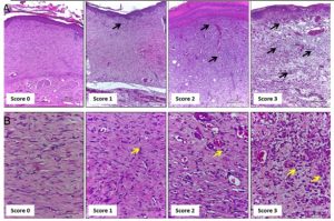
Figure 01: Representative photomicrographs of sections stained with H & E showing the inflammation score
Score 0: skin; Score 1: the wound of Days 21 and 14; Score 2: on Day 7, score 3: on Day 3. Inflammatory cells indicated by black and yellow arrows at different concentrations according to the healing time. A. Scale bars: 200 μm B. Scale bars: 50 μm. The amount of extracellular matrix deposited, and the thickness of collagen fibers in H and E-stained sections were evaluated using the 10x objective.
Table 02: Morphological scores of extracellular matrix deposition in H and E-stained histological sections of excisional rat wounds
| Score | Parameter |
| 0 | WHOLE SKIN–whole extracellular matrix |
| 1 | DISCRETE–incomplete presence of extracellular matrix (identified by many fibroblasts, and thin collagen fibers) |
| 2 | MODERATE–presence of extracellular matrix in the whole wound area (identified by many fibroblasts, and thin collagen fibers) |
| 3 | HIGH–presence of extracellular matrix in the whole wound area (identified by few fibroblasts, and thick collagen fibers) |
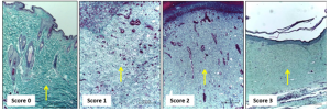
Figure 02: Representative photomicrographs of Gomori’s trichrome-stained sections showing the score of extracellular matrix deposition
Score 0: skin; Score 1: on Day 3; Score 2: on Day 7, Score 3: on Day 14 and Day 21. Scale bars: 200 μm. Evaluate the amount of blood vessels in the new tissue formed in HE-stained sections using the 40x objective.
Table 03: Morphological scores of vascularization in H&E-stained histological sections of excisional rat’s wounds
| Score | Parameter |
| 0 | WHOLE SKIN–normal vascularization |
| 1 | DISCRETE VASCULAR FORMATION |
| 2 | MODERATE VASCULAR FORMATION |
| 3 | HIGH VASCULAR FORMATION |
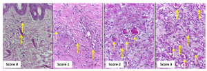
Figure 03: Representative photomicrographs of H & E-stained sections showing the score of vascularization
Score 0: whole skin; Scores 1: on Day 21; Score 2: on Day 14, Score 3: on Days 3 and 7. Scale bars: 50 μm. Department Committee ethics approval was granted for this project from Agriculture and Forestry University in Chitwan, Nepal, with the consent of a faculty of veterinary surgery and pharmacology. The National Ethical Guidelines for Health Research in Nepal and Standard Operating Procedure 2011 and the Ethical Guidelines for the use and care of animals in health research in NEPAL 2005 were followed in all of the study’s procedures. All operations were performed under anesthetic, and every attempt was made to lessen the pain.
- RESULT & DISCUSSION
Evaluation of wound contraction percentage and period of epithelialization Using procedures, the percentage of wound contraction was estimated, and the analysis was displayed graphically.
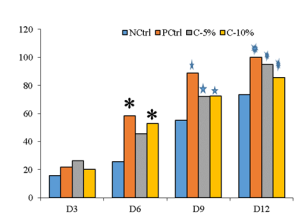
Figure 04: Wound contraction percentage of Cissus quadrangularis in rats at AFU
Following three days after wounding, a significant increase in the percentage of wound contraction was observed across the board. Six days after the injury, the 5% concentration exhibited slower healing, but on days nine and twelve, healing considerably and proportionately increased across all groups. After three days’ post-injury, the contraction rate significantly increased, reaching its peak 12 days after the operation. Epithelialization lasted for the control, standard, 10%, and 5% treatment groups, respectively, for 25, 18, 15, and 15 days. By using a digital automatic blood analyzer to evaluate a blood sample, blood parameters are discovered. When compared to the vehicle-treated group, we observed a decrease in the granulocyte and RBC counts of the lesions treated with plant extract and regular ointment.
Table 04: Blood parameter of samples
| Days after wounding | |||||||||||||||||
| Hematological parameter | 3 | 6 | 9 | 12 | |||||||||||||
| NC | 5% | 10% | PC | NC | 5% | 10% | PC | NC | 5% | 10% | PC | NC | 5% | 10% | PC | ||
| Platelets | 1082 | 1082 | 1021 | 1021 | 763 | 1021 | 800 | 779 | 513 | 1001 | 228↓ | 604↓ | 44↓ | 307↓ | 204↓ | 65↓ | |
| GRAN# | 0.4 | 0.8 | 0.02 | 1 | 0.8 | 3.7 | 2.5 | 2.5↑ | 0.7 | 1 | 2.5 | 1.9 | 0.7 | 0.8 | 0.5 | 0.7 | |
| RBC | 1.8 | 3.24↓ | 2.87↓ | 3.41↓ | 6.3↓ | 5.84↓ | 6.3 | 6.30↓ | 5.84↓ | 5.64↓ | 6.3↓ | 6.08 | 5.17↓ | 4.46↓ | 5.73↓ | 4.90↓ | |
| WBC | 1.8 | 5.9 | 3.8 | 2.3↓ | 6.9↑ | 10.9↑ | 7.4↑ | 9.8 | 7.4↑ | 6.3 | 7.4 | 7.4↑ | 6 | 6 | 5.5 | 4.3 | |
| LYM# | 1 | 4.4 | 2.6 | 3.2 | 4.0↑ | 4.7 | 4.5 | 4.2 | 4.5 | 4.4 | 4.5 | 4.7 | 7.1↑ | 4.5 | 7.1↑ | ||
| HBG | 4.8↑ | 17.0↑ | 19.3↑ | 17.7↑ | 16.4↑ | 19.9↑ | 18.3 | 18.3↑ | 18.3↑ | 15.1↑ | 18.0↑ | 0.9↓ | 19.9↑ | 14.1↑ | 6.9↑ | 19.6↑ | |
| HCT | 11.3↓ | 37.1 | 41.1 | 34.9 | 39.2 | 39.4 | 17.5↓ | 41.1 | 41.1 | 36.3 | 38.7 | 2.8↓ | 39.4 | 32.5↓ | 41.1 | 35.2 | |
Table 05: Morphological (histopathological) scores obtained by analyzing the H & E-stained tissue for each parameter according to bio-protocol 9(13): e3285 [3].
| Days after wounding | ||||||||||||||||
| Histopathological Parameters | 3 days | 6 days | 9 days | 12 days | ||||||||||||
| NC | 5% | 10% | PC | NC | 5% | 10% | PC | NC | 5% | 10% | PC | NC | 5% | 10% | PC | |
| Inflammation | 3 | 1 | 1 | 1 | 2 | 1 | 1 | 1 | 2 | 0 | 0 | 0 | 1 | 0 | 0 | 0 |
| Extracellular matrix deposition | 1 | 1 | 1 | 1 | 1 | 2 | 2 | 2 | 2 | 2 | 2 | 2 | 2 | 3 | 3 | 3 |
| Angiogenesis | 1 | 1 | 1 | 1 | 2 | 3 | 3 | 3 | 3 | 2 | 2 | 2 | 2 | 1 | 0 | 0 |
Inflammatory cells accumulation is reduced in plant and standard treatment group. I observed a reduction in the inflammatory cells accumulation of wounds from plant treated and standard ointment treated group, when compared to Vehicle treated group, as analyzed histologically Figure 05 and 06.
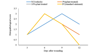
Figure 05: Inflammatory cell pattern after wound creation.
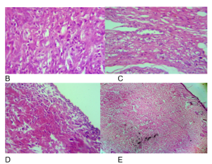
Figure 06: Inflammatory cells accumulation is reduced in wounds from plant-treated and standard antibiotic ointments compared to vehicle treated group. (A) histopathological score of different treatment groups days after wounding. Representative photomicrographs of H & E-stained sections of different histopathological score (B-3, C-2, D-1 and E-0).
Extracellular matrix deposition is increased in plant and standard treatment group. I observed increase in the Extracellular matrix deposition of wounds from plant treated and standard ointment treated group, when compared to Vehicle treated group, as analyzed histologically (Figure 07 & 08).
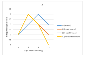
Figure 07: Extracellular matrix cell deposition on wound sites
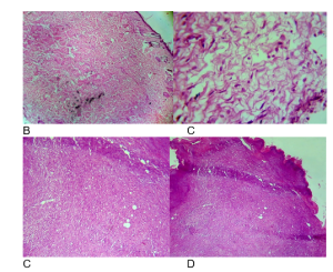
Figure 08: Extracellular matrix deposition is increased in wounds from plant-treated and standard antibiotic ointments compared to vehicle treated group. (A) histopathological score of different treatment groups, days after wounding. Representative photomicrographs of H& E-stained sections of different histopathological score (B-3, C-2, D-1 and E-0)
Angiogenesis is reduced in plant and standard treatment group. I observed a reduction in angiogenesis of wounds from plant treated and standard ointment treated group, when compared to Vehicle treated group, as analyzed histologically (Figure 9 and 10).
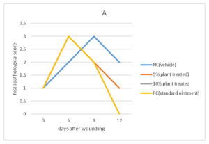
Figure 09: Graphical representation of scoring of wound healing on the basis of angiogenesis.
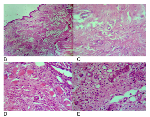
Figure 10: angiogenesis is reduced in wounds from plant-treated and standard antibiotic ointments compared to vehicle treated group. (A) histopathological score of different treatment groups, days after wounding. Representative photomicrographs of H & E-stained sections of different histopathological score (B-3, C-2, D-1 and E-0).
Alkaloids, Flavonoids, Terpenoids, Glycosides, and Saponins were discovered in the 80% methanolic extract of air-dried shoots of Cissus quadrangularis after phytochemical investigation. The goal of the current study was to analyze the phytochemical composition of Cissus quadrangularis and assess its ability to heal wounds in albino rats. The results of this property’s evaluation indicate that the plant has a statistically significant influence on wound healing when compared to the control group. The 5% dosage of Cissus quadrangularis in this test performed significantly better than the 10% extracts. The excision wound model’s findings show that, with the exception of the control group, there was no discernible increase in wound contraction after three days of sampling. Following three days of healing, the findings of the subsequent days show a considerable rise (p 0.001). The outcome was also confirmed by hematological and histopathological analysis. These findings concur with those of [17] for epithelialization period. In contrast to [18], this study used Cissus quadrangularis ethosomal gels, which have the ability to speed up wound healing as compared to the gold standard (mebo ointment) in 12–14 days. This variety could result from different plant extract percentages, rat meals, the timing of the research, etc. Similar to what is stated in this article, [12] similarly found that extracts dramatically accelerated wound contraction on the 12th day (p 0.001) compared to the control group. An author [19] found that applying an ointment containing 2% (w/w) methanolic and 2% (w/w) total aqueous extract to a wound exhibited significant (P 0.001) wound healing activity. In the case of the excision wound model, the duration of epithelization and the contraction of the wound were also examined. Crude methanolic extracts of C. quadrangularis, A. multiflorum, and E. abyssinica (leaf and bark separately) were utilized in the tests to determine the wound-healing abilities of the freshly prepared ointments. concluded that during a 15-day period, ointments of C. quadrangularis and A. multiflorum caused wound alterations and healing that was similar to the positive control [20]. An author carried out a similar study, but there were distinctions. 18 goats of various ages and sexes that had wounds were split into three groups of six each. In the control group, the wounds were cleaned with normal saline; however, in the other two groups, the wounds were then treated with either a 5.0 or 7.5% methanolic extract ointment of C. quadrangularis. In each group, observations were made after 0, 4, 7, and 14 days. Complete healing was attained in the majority of cases by the 14th day thanks to the prepared ointments, which had a noticeable impact on the rate of healing [21]. The outcome of the phytochemical study of the 80% methanolic extract of the air-dried shoot of Cissus quadrangularis in this experiment was comparable to that of [22]. On this shoot, tannins and alkaloids are found. Alkaloids, sugars, glycosides, saponins, sterols, phenolic compounds, and flavonoids were all observed by [23] to be present in plants. The different substances found in the same plant may be caused, in part, by differences in the type of extract utilized, the plant parts used, the age of the plant, or the analytical technique. The Phytochemical study of Cissus quadrangularis have shown the presence of various versatile constituents such as flavonoids, triterpenoids, Vitamin C, stilbene derivatives and many others, e.g. resveratrol, piceatannol, pallidol, perthenocissin and phytosterols. Out of which ascorbic acid, triterpene, β-sitosterol, ketosteroid, two asymmetrical tetracyclic triterpenoids and calcium were identified as major constituents of this plant [23,24,25].
- CONCLUSION
The goal of the current study was to analyze the phytochemical composition of Cissus quadrangularis and assess its ability to heal wounds in albino rats. The results of this property’s evaluation indicate that the plant has a statistically significant influence on wound healing when compared to the control group. The 5% dosage of Cissus quadrangularis in this test performed significantly better than the 10% extracts. The outcome of the phytochemical study of the 80% methanolic extract of the air-dried shoot of Cissus quadrangularis in this experiment found shoot, tannins and alkaloids. Alkaloids, sugars, glycosides, saponins, sterols, phenolic compounds, and flavonoids were all observed to be present in plants. The different substances found in the same plant may be caused, in part, by differences in the type of extract utilized, the plant parts used, the age of the plant, or the analytical technique. The excision wound model’s findings show that, with the exception of the control group, there was no discernible increase in wound contraction after three days of sampling. Following three days of healing, the findings of the subsequent days show a considerable rise (p 0.001). The outcome was also confirmed by hematological and histopathological analysis. After three days of therapy, the Cissus quadrangularis extract’s wound contraction % was much higher than that of the control group, and a representative sample’s hematological and histopathological analysis amply supported this conclusion. This study demonstrated the lack of wound-healing abilities of Cissus quadrangularis. This kind of research may aid in examining the potential creation of future generation medicines that are very effective but have less adverse effects. Since AMR is currently a problem with all antibiotics, this kind of medicine composition can also lessen the risk of AMR. Because the wound-healing abilities of Cissus quadrangularis have never been studied and have been passed down verbally over generations, they can be a very useful tool for drug formulation companies and researchers alike.
REFERENCES
- Bharti, M., Borane, K., & Singhasiya, A. (2014). Evaluation of wound healing activity of Cissus quadrangularis. World Journal of Pharmacy and Pharmaceutical Sciences, 3(6), 822–824.
- Bhat, M. A., & Aher, V. D. (2014). Clinical evaluation of Cissus quadrangularis on wound healing in caprine (Capra hircus). SKUAST Journal of Research, 13(2), 92–96.
- de Moura Estevão, L. R., Cassini-Vieira, P., Leite, A. G. B., de Carvalho Bulhões, A. A. V., da Silva Barcelos, L., & Evêncio-Neto, J. (2019). Morphological evaluation of wound healing events in the excisional wound healing model in rats. Bio-Protocol, 9(13), e3285. https://doi.org/10.21769/BioProtoc.3285
- Deodhar, A. K., & Rana, R. E. (1997). Surgical physiology of wound healing: A review. Journal of Postgraduate Medicine, 43(2), 52–56.
- Emami-Razavi, S. H., Esmaeili, N., Forouzannia, S. K., Amanpour, S., Rabbani, S., Alizadeh, A. M., & Mohagheghi, M. A. (2006). Effect of bentonite on skin wound healing: Experimental study in the rat model. Acta Medica Iranica, 44(4), 235–240.
- Friedman, A. (2011). Wound healing: From basic science to clinical practice and beyond. Journal of Drugs in Dermatology, 10(4), 427–433.
- Karodi, R., Jadhav, M. M., Rub, R. A., & Bafna, A. (2009). Evaluation of the wound healing activity of a crude extract of Rubia cordifolia (Indian madder) in mice. International Journal of Applied Research in Natural Products, 2(2), 12–18.
- Kumar, R., Katoch, S. S., & Sharma, S. (2006). Beta-adrenoceptor agonist treatment reverses denervation atrophy with augmentation of collagen proliferation in denervated mice gastrocnemius muscle. Indian Journal of Experimental Biology, 44(5), 371–376.
- Kunwar, R. M., Uprety, Y., Burlakoti, C., Chowdhary, C. L., & Bussmann, R. W. (2009). Indigenous use and ethnopharmacology of medicinal plants in far-west Nepal. Ethnobotany Research and Applications, 7, 005–028.
- Mohanty, A., Sahu, P. K., & Das, C. (2010). Wound healing activities of methanolic extract of Cissus quadrangularis on albino rats. International Journal of Drug Formulation and Research, 1, 176–183. (Verify page range).
- Marume, A., Matope, G., Katsande, S., Khoza, S., Mutingwende, I., Mduluza, T., Munodawafa-Taderera, T., & Ndhlala, A. R. (2017). Wound healing properties of selected plants used in ethnoveterinary medicine. Frontiers in Pharmacology, 8, 544. https://doi.org/10.3389/fphar.2017.00544
- Murthy, S., Gautam, M. K., Goel, S., Purohit, V., Sharma, H., & Goel, R. K. (2013). Evaluation of in vivo wound healing activity of Bacopa monnieri on different wound models in rats. BioMed Research International, 2013, Article 972028. https://doi.org/10.1155/2013/972028
- Siddique, A., & Mittapaly, S. (2017). Pharmacological evaluation of Cissus quadrangularis herbal gel in wound healing activity. International Journal of Research in Ayurveda and Pharmacy, 8(5), 65–68. https://doi.org/10.7897/2277-4343.085259
- Senthil Kumar, M., Sripriya, R., Vijaya Raghavan, H., & Sehgal, P. K. (2006). Wound healing potential of Cassia fistula on infected albino rat model. Journal of Surgical Research, 131(2), 283–289. https://doi.org/10.1016/j.jss.2005.08.025
- Zhong, S. P., Zhang, Y. Z., & Lim, C. T. (2010). Tissue scaffolds for skin wound healing and dermal reconstruction. Wiley Interdisciplinary Reviews: Nanomedicine and Nanobiotechnology, 2(5), 510–525. https://doi.org/10.1002/wnan.100
- Ramos-e-Silva, M., & Ribeiro de Castro, M. C. (2002). New dressings, including tissue-engineered living skin. Clinics in Dermatology, 20(6), 715–723. https://doi.org/10.1016/S0738-081X(02)00298-5
- Dhivya, S., Padma, V. V., & Santhini, E. (2015). Wound dressings—A review. BioMedicine, 5(4), 22. https://doi.org/10.7603/s40681-015-0022-9
- Lai, J. Y. (2012). Biocompatibility of genipin and glutaraldehyde cross-linked chitosan materials in the anterior chamber of the eye. International Journal of Molecular Sciences, 13(9), 10970–10984. https://doi.org/10.3390/ijms130910970
- Mutlu, G. (2019). Preparation and characterization of active agent loaded nanofibrillar structures for tissue regeneration (Master’s thesis). (Institution name required for complete APA citation.)
- Mogoșanu, G. D., & Grumezescu, A. M. (2014). Natural and synthetic polymers for wounds and burns dressing. International Journal of Pharmaceutics, 463(2), 127–136. https://doi.org/10.1016/j.ijpharm.2013.12.015
- Jainu, M., & Devi, C. S. S. (2003). Potent antiulcerogenic activity of Cissus quadrangularis on aspirin-induced gastric ulcer by its antioxidative mechanism. Journal of Clinical Biochemistry and Nutrition, 34, 43–47.
- Jainu, M., & Devi, C. S. S. (2003). Potent antiulcerogenic activity of methanolic extract of Cissus quadrangularis by antioxidative mechanism. Journal of Clinical Biochemistry and Nutrition, 34, 43–47.
- Enechi, O. C., & Odonwodo, I. (2003). An assessment of the phytochemical and nutrient composition of the pulverized root of Cissus quadrangularis. BioResearch, 1(1), 63–68.
- Shirley, D. A., & Sen, S. P. (1966). High-resolution X-ray photoemission studies on the active constituents of Cissus quadrangularis. Current Science, 35, 317.
- Rao, K. N. V., Bhongiri, B., Thirupathaiah, A., Mamata, M., & Illendula, S. (2026). Cissus Quadrangularis L-A Comprehensive Multidisciplinary Review. Journal of Applied Pharmaceutical Sciences and Research, 9(03), 1-13.
Publication History
Submitted: October 31, 2024
Accepted: November 22, 2024
Published: March 31, 2025
Identification
D-0595
DOI
https://doi.org/10.71017/djnsi.4.5.d-0595
Citation
Binu Kumari Shah, Dipendra Kumar Sah & Manoj Shah (2025). Evolution Of the Wound Healing Activity of Cissus Quadrangularis on Excision Wound Models in Albino Rats. Dinkum Journal of Natural & Scientific Innovations, 4(04):278-290.
Copyright
© 2025 The Author(s).