

Publication History
Submitted: October 15, 2025
Accepted: November 11, 2025
Published: November 30, 2025
Identification
D-0547
DOI
https://doi.org/10.71017/djmi.4.11.d-0547
Citation
Rashmi Burlakoti, Ram Kumar Ghimire, Swoyam Prakash Pandit, Om Biju Pant, Sagar Nepal & Neelam Maharjan (2025). Frequency of Malignant Thyroid Nodule on Fine Needle Aspiration Cytology on Patient Found to be Positive on Ultrasonography Dinkum Journal of Medical Innovations, 4(11):745-754.
Copyright
© 2025 The Author(s).

745-754
Frequency of Malignant Thyroid Nodule on Fine Needle Aspiration Cytology on Patient Found to be Positive on UltrasonographyOriginal Article
Rashmi Burlakoti 1*, Ram Kumar Ghimire 2, Swoyam Prakash Pandit 3, Om Biju Pant 4, Sagar Nepal 5, Neelam Maharjan 6
- College of Physicians and Surgeons (CPSP), Pakistan
- Tribhuwan University, Institute of Medicine, Kathmandu Nepal.
- Tribhuwan University, Institute of Medicine, Kathmandu Nepal.
- Tribhuwan University, Institute of Medicine, Kathmandu Nepal.
- Philippine Department of Health, Jose R. Reyes Memorial Medical Centre, Manila, Philippines
- College of Physicians and Surgeons (CPSP), Pakistan
* Correspondence: ras.burlakoti.rb@gmail.com
Abstract: Thyroid nodules are a very common clinical finding, which presents in various forms of endocrinological disorder such as hypothyroidism, hyperthyroidism or as a palpable lump or goiter. Some of them may be asymptomatic. Thyroid disease being a common health problem in our country, a study evaluating the frequency of malignant thyroid disease was essentially necessary. Therefore, we aimed to evaluate the frequency of malignant thyroid nodule initially by ultrasonography (USG) followed by USG guided fine needle aspiration cytology (FNAC). This study determined the frequency of malignant thyroid nodules on FNAC on nodules that are found to have malignant features in USG. Sonographic malignant features corelated with FNAC result. This study was prospective cross-sectional and was conducted in Department of Radiodiagnosis of Nepal Mediciti Hospital, Lalitpur. Duration of Study was 6 months. One hundred fifty-four patients having thyroid nodule with at least one suspicious malignant sonographic feature among hypoechoic, microcalcification, taller than wider and irregular margin were included in the study. These features were recorded followed by FNAC. A total of 154 patients with at least one suspicious malignant were included in the study. In cytological examinations, 80 nodules were malignant, and 74 nodules were benign. Among four suspicious malignant sonographic features, microcalcification was associated with higher frequency in malignant nodules. Ultrasonography is a valuable tool in differentiating malignant from benign thyroid lesions. Microcalcification is a strong predictor of malignancy.
Keywords: thyroid nodule, ultrasonography, fine needle aspiration cytology
1.INTRODUCTION
Thyroid nodules are a very common clinical finding, which presents in various forms of endocrinological disorder such as hypothyroidism, hyperthyroidism or as a palpable lump or goiter. Some of them may be asymptomatic [1]. The thyroid gland originates at the level of foramen cecum in the base of the tongue between the first and second pharyngeal pouches [2]. At third week of gestation, thyroid diverticulum is formed by the proliferation of endodermal cells of the primitive pharynx. At 5th week, thyroid diverticulum descends along the midline anterior to the structures forming the larynx. It then divides into right and left lobe. Thyroid remains attached to the base of the tongue via the thyroglossal duct. Initially, thyroid gland is hollow. Later on, during migration, it solidifies forming the follicular cells [3]. Also, during the fifth week, the paired ultimobranchial bodies arise from the fourth/fifth pharyngeal pouches. They provide the parafollicular C cells producing calcitonin [4]. The ultimobranchial bodies fuse with the superior dorsolateral aspect of the developing thyroid, forming Zuckerkandl’s tubercle [5]. Thyroid gland reaches its destination in the neck by seventh week of gestation. Normally, the thyroglossal duct degenerates by the tenth week of gestation. Incomplete obliteration of the duct can lead to thyroglossal duct cysts, lingual thyroid, or a pyramidal lobe. Thyroid gland becomes functionally mature by the twelfth gestational week [1]. The prevalence of thyroid nodule worldwide has been reported to be 2-6% with palpation, 19-35% with Ultrasonography (USG), and 8%-65% on autopsy. Most of the thyroid nodules are benign, with malignant having 5% cases. The incidence of malignant thyroid nodule in Nepal is 11.21% [2]. The reported frequency of malignant thyroid nodule on ultrasonography (USG) is 20% [3]. The prevalence of sonographically detected nodules that cannot be palpated is up to 50-60% in individuals older than 60 [4]. Therefore, it is a challenge for the clinicians to identify the nodules that have high probability of being malignant. The use of high-frequency sonographic imaging in evaluation of the thyroid gland has led to the detection of a large number of non-palpable thyroid nodules in the general population. USG was first used to diagnose thyroid nodules in 1967 by Fujimoto [5]. Current methods of USG permit real-time identification of structures as small as 2 mm in diameter, thereby allowing the visualization of very small nodules. Doppler techniques can be added to differentiate cystic and vascular structures [6]. Many times, thyroid nodules come to clinical attention when noted by the patient or as an incidental finding during routine physical examination or radiologic procedure, such as carotid ultrasonography or neck computed tomography (CT) [7]. History and physical examinations are mandatory in a patient with thyroid nodule. However, in a small percentage of patient’s symptoms are confusing. Good history taking and examination in the assessment of thyroid nodules. Physical examination factors associated with increased likelihood of malignancy include firmness of the nodule, rapid growth, fixation to adjacent structures, vocal cord paralysis, and enlarged regional lymph nodes [8]. Most patients with benign or malignant thyroid nodules have euthyroid status. Despite this, serum thyroid stimulating hormone (TSH) measurement is recommended in all patients presenting with a nodule [7,8]. In the case of low serum TSH, a radionuclide thyroid scintigraphy (99Tc or 123I) should be obtained to document whether the nodule is functioning (hot) or not. Functioning nodules rarely harbor malignancy. Hence, in the event of a functioning nodule, some authors suggest that no additional cytological evaluation is necessary. If overt or subclinical hyperthyroidism is present, additional evaluation of the hyperthyroidism is required. In cases of normal or high serum TSH, diagnostic thyroid USG should be performed [9]. Even if the TSH is high, Fine needle aspiration cytology (FNAC) is recommended, as the rate of malignancy in nodules within thyroid glands with Hashimoto’s thyroiditis (high TSH) is similar to nodules within thyroid glands without Hashimoto’s thyroiditis (normal TSH). If TSH is high, antithyroid antibodies (anti-TPO, anti-TG) should also be obtained to confirm the diagnosis of Hashimoto’s thyroiditis [10]. It has recently been reported that the risk of malignancy in a thyroid nodule increases proportionally to serum TSH concentrations at presentation, even within the normal range, and thus TSH was proposed as a novel independent predictor of the presence of thyroid malignancy [11]. After excluding the nodules with low serum TSH levels, recently published evidence-based guidelines from the American Thyroid Association and the American Association of Clinical Endocrinologists recommend a diagnostic thyroid ultrasound for patients with palpable thyroid nodules [9,12]. The rationale for this includes: Confirmation of a sonographically identifiable nodule corresponding to the palpable abnormality. Detection of additional nonpalpable nodules for which FNA may be indicated. Determination of accuracy of FNAC by palpation. Identification of the sonographic characteristics of the thyroid nodule. Even though several studies have been designed to evaluate whether USG can be used in the differentiation of benign and malignant thyroid nodules, diagnostic USG findings of malignancy do not exist. Certain USG parameters are useful in the clinical decision, and they include margin, echotexture, echogenicity, calcification, internal vasculature, etc. Fine needle aspiration cytology (FNAC) is considered the most reliable diagnostic test for the evaluation of thyroid nodules. It has a low rate of complications when ultrasound guidance is used [13]. This study focused on different parameters in USG in evaluating thyroid nodules for malignancy, its accuracy in diagnosing malignant nodules, and its correlation with FNAC. Nevertheless, there is significant uncertainty surrounding the diagnostic accuracy of several of the features analyzed during the sonographic evaluation of thyroid nodules. This study helped to acknowledge the malignant features of thyroid nodules on USG and whether the findings correlate with the histopathological report or not. A better understanding of the US features predictive of malignancy or benign disease may avoid costly confirmatory testing and have a large impact on both guideline recommendations and clinical practice.
2.MATERIALS AND METHODS
The study was conducted in Department of Radio diagnosis of Nepal Mediciti Hospital, Bhaisepati, Lalitpur. The case collection and study were done for 6 months after approval of synopsis. Taking frequency of thyroid nodule on FNAC which were malignant on USG as 57.9% [14], 95% level of confidence and absolute precision of 7.3%, total sample size would be around 154. Consecutive sampling was done. This study was prospective cross-sectional study. Data collection was done in the Department of Radiology, Nepal Mediciti Hospital. Patient referred from various departments were scanned. Evaluation of thyroid gland was done by high- resolution real-time ultrasonography in Samsung ultrasound machine by linear probe of LA 3-16 MHz. The patient lying supine and the neck in a slightly hyper-extended position to fully expose the anterior neck. A semi-erect position was acceptable if the patient was unable to tolerate the preferred posture. The nodules were characterized by echogenicity, calcification shape and margin. Hypoechoic, microcalcification, taller than wider and irregular margin were considered as four suspicious malignant features. If any one of the malignant features were noted, then the patient was included in the study and subjected to FNAC. USG guided FNAC was done by using non aspiration technique with 25 G spinal needle. At least 2 passes were done. Needle was directed to the site with suspicious features. 6 slides were prepared,3 air dried and 3 alcohols soaked. If multiple nodules were present, the most suspicious one was selected for FNAC. Adequacy of the sample was determined by cytopathological technologist immediately after fine needle aspiration at procedure room. Data were collected using a preformed proforma and statistical analysis was done by IBM, Statistical Package for Social Sciences (SPSS) version 22.0. Descriptive analysis was done to calculate frequencies and percentages of qualitative variables like sex, single/multiple nodules and malignant thyroid nodule on FNAC. Mean and standard deviation were calculated for quantitative variable like age, duration of swelling or symptoms. Effect modifiers like age, gender and duration of swelling or symptoms, single/multiple nodules were stratified to see the effect of these on outcome. Post stratification chi square test was applied. P value less than or equal to 0.05 was considered as significant.
3.RESULTS AND DISCUSSION
A total of 156 patients were included in the study. FNAC of 2 nodules were inconclusive and were excluded from the study making sample size of 154. Out of the 154 nodules, FNAC diagnosis of malignant nodule was made in 80 (51.9%) and diagnosis of benign nodules was made in 74 (48.1%). Out of the benign nodules, 4 cases were thyroiditis. Saphiro Wilk Test was significant (p value of 0.09) demonstrating the age distribution follows a normal distribution. Among age ranged from 20-75 years, the mean age of the population was 46.27+/-14.217, with median of 46 (IQR 21). Our population predominantly consisted of females. 72.08% were females and 27.92% were male (Table 01). Among 154 population, 122(79.2%) presented with single nodules and 32(20.8%) presented with multiple nodules (Table 02). The mean duration of symptom was 28.44+/-39.53 weeks with median of 12 weeks and interquartile range of 34 weeks (Table 03). 29 cases had symptoms less than 4 weeks. 4 of the cases were detected incidentally during carotid Doppler study (Table 04). Hypoechoic nature was the most common suspicious USG feature encountered and taller than wide shape was the least common USG feature encountered in the study (Table 05). Only one suspicious USG feature was present in 90(58.4%), 2 suspicious features were present in 50(32.5%), 3 suspicious features were present in 11(7.1%), and all 4 suspicious feature was present in 3(1.9%) nodules (Table 06). FNAC of these nodules revealed malignancy in 80 (51.95%) cases with 49.5% of females (55/111) and 58.1 % of males (25/43). Malignancy was seen in 64 and benignity were seen in 58 of the solitary nodules. However, no significant difference noted in benignity and malignancy in multiple nodules (Table 07). Table 8 shows that out of 80 malignant proven cases, hypoechoic nature was seen in 60 nodules, microcalcification was seen in 47 cases, taller than wider shape was seen in 19 cases and irregular margin was seen in 20 cases. Among 74 benign nodules, hypoechogenecity was the most encountered feature. It was found that presence of microcalcification is a good predictor of malignancy with p value <0.001.
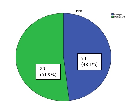
Figure 01: Pie chart showing FNAC diagnosis of thyroid nodules
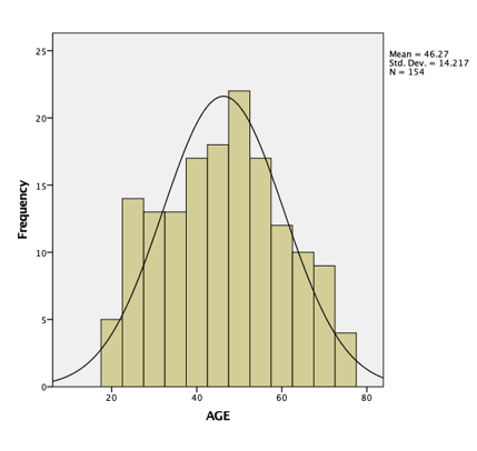
Figure 02: Histogram showing normal age distribution and mean age.
Table 01: Patient distribution based on sex
| Sex | Frequency and percentage |
| Male | 43 (27.92 %) |
| Female | 111 (72.08%) |
Table 02: Frequency and percentage of single and multiple nodules
| Number of nodules | Frequency | Percentage |
| Single | 122 | 79.2% |
| Multiple | 32 | 20.8% |
Table 03: Duration of symptoms
| Duration of symptoms | Weeks |
| Mean | 28.44 |
| S. D | 39.53 |
| Median | 12 |
| Interquartile range | 34 |
Table 04: Duration of symptoms and number of cases
| cases Duration of symptoms Number of cases |
| <4 weeks 29 |
| >4 weeks 121 |
| Incidental finding 4 |
Table 05: Malignant ultrasonography features of thyroid nodules
| Features | Number (percentage) N=154 |
| Hypoechoic nature | 105(68.2) |
| Presence of microcalcifications | 69(44.8)
|
| Irregular Margin | 32(20.8) |
| Taller than wide shape (Nonparallel orientation) | 29(18.8)
|
Table 06: Number of malignant features in thyroid nodules
| Number of malignant features | Number of cases |
| One | 90 (58.4%) |
| Two | 50 (32.5%) |
| Three | 11 (7.1%) |
| Four | 3 (1.9%) |
Table 07: Correlation between demographics of population with histopathological examination.
| Demographics | Benign
N=74 |
Malignant
N=80 |
p-value | |
| Age: | ||||
| Mean | 50.92 | 44.51 | 0.01* | |
| S. D | 15.77 | 15.01 | ||
| Sex: | ||||
| Male | 18 | 25 | 0.37# | |
| Female | 56 | 55 | ||
| Duration in | ||||
| Weeks | 0.14* | |||
| Mean | 23.64 | 32.95 | ||
| S. D | 0.57 | 46.12 | ||
| Number of | ||||
| lesions | 0.80# | |||
| Single | 58 | 64 | ||
| Multiple | 16 | 16 | ||
Table 08: Correlation of suspicious USG features with cytopathological findings
| USG Features | Benign
N=74 |
Malignant
N=80 |
p-value |
| Hypoechoic appearance | 45 | 60 | 0.059# |
| Presence of microcalcification | 22 | 47 | <0.001# |
| Taller than wider shape (Nonparallel orientation) | 10 | 19 | 0.10# |
| Irregular margin | 12 | 20 | 0.23# |
Thyroid ultrasound is an exquisitely sensitive technique for the detection of thyroid nodules and is able to image nodules as small as 2–3 mm. The prevalence of sonographically detected nodules that cannot be palpated is up to 50%–60% in individuals older than 60 [15]. Therefore, the challenges to the clinician are the identification of those nodules that have a higher probability of being clinically relevant malignancies so that these can be targeted for fine-needle aspiration biopsy, and the recognition of those that may undergo sonographic surveillance. Several US characteristics have been studied as potential predictors of thyroid malignancy but there is also overlap in their appearances [16]. Because of the inconsistent predictive value of US features, FNA and cytopathologic evaluation of a thyroid nodule are usually required before a patient undergoes surgical resection for a possible thyroid malignancy [17]. The widespread use of FNA and cytopathologic analysis has improved the detection of thyroid cancer and has led to a decreased frequency of thyroid surgery [18]. This study focused at correlation of suspicious ultrasound characteristics of thyroid nodules with FNAC findings and hence determines the importance of each characteristic in determining the malignancy of the thyroid nodule. In this study, a total of 154 patients from different parts of country were included. The mean age group was 47.59+/-15.66 years. Similar were the findings in the study [19] with mean age was 41.2±15 years. In this study, malignant thyroid nodule was seen more commonly on female that male in the ratio of 2.2:1. The observations are almost similar to the female: male ratio of 3.4:1 in the studies by [20] In the present study, 49.5% females and 58.1 % males had malignant nodules suggesting that thyroid nodules though more prevalent in females, malignancy however was observed more among males. Similar were the findings in the study by [21]. Hypoechogenecity was the most common encountered US features. Out of 105 hypoechoic nodules, 60 were found to be malignant nodules and 45 were found to be benign nodules. Though both benign and malignant nodules can appear hypoechoic, it was more prevalent among malignant. Study done by [22]. An author [23] result was that echogenicity did not show any significant difference between benign and malignant nodules. Study done by [24] found that association of markedly hypo echogenicity was significantly different between benign and malignant nodules (p<0.0001). Most of malignant thyroid nodule was markedly hypoechogenic in comparison to the benign nodules. In this study, out of 69 nodules having microcalcification in USG, 47 were malignant and 22 were benign nodules. Microcalcification was more common in malignant nodules, and it has been found in this study that microcalcification is a significant predictor of malignancy (p value < 0.001). An author [25] in his study identified intrinsic calcification as the only statistically significant predictor of malignancy (35.3% sensitive and 94.4% specific; P < .005). In study done by [26], they have stated that microcalcification, although uncommon, can be considered nearly specific for malignancy. Multilogistic regression analysis done in study carried out by [27] showed that microcalcification was a significant independent factor for predicting malignancy (p = 0.0022). Taller than wider was also seen in 19 of the malignant thyroid nodules and 10 of the benign nodules. Study conducted by [28] also suggest a taller than-wide shape is very specific for differentiating malignant thyroid nodules from benign ones. But having low sensitivity, it can be used as ancillary finding for thyroid malignancy. Out of 32 nodules with irregular margin, 20 nodules were found to be malignant and 12 were found to be benign. Similar were the result in study conducted by [29]. Their study results depicted sensitivity, specificity, PPV, NPV and diagnostic accuracy of 12.5%, 94.74%, 42.85%, 77.42% and 75% respectively.
4.CONCLUSION
Ultrasonography is a valuable and effective imaging modality for the evaluation and risk stratification of thyroid nodules. In this study, approximately half of the thyroid nodules exhibiting at least one suspicious malignant sonographic feature were confirmed as malignant on FNAC. Among the evaluated ultrasound characteristics, microcalcification demonstrated the strongest association with malignancy and emerged as a significant predictor of malignant thyroid nodules. In contrast, other suspicious sonographic features, including hypoechogenicity, irregular margins, and a taller-than-wide shape, were observed in both benign and malignant nodules, thereby reducing their individual diagnostic specificity. These findings underscore the importance of careful sonographic assessment in the identification of thyroid nodules at increased risk of malignancy. Familiarity with malignant ultrasound features, particularly microcalcifications, can enhance diagnostic accuracy and assist clinicians and radiologists in selecting appropriate candidates for FNAC. The integration of ultrasonographic findings with cytological evaluation provides a reliable approach to the diagnosis and management of thyroid nodules, facilitating early detection of thyroid malignancies while potentially reducing unnecessary invasive procedures in low-risk cases. Ultimately, this approach supports evidence-based clinical decision-making and contributes to improved patient outcomes.
REFERENCE:
- Sandler MP, Patton JA, McCook BM. Multimodality imaging of the thyroid gland. Baillieres Clin Endocrinol Metab. 1989 May;3(1):89–119.
- Acharya Barsha, Smriti Karki, Anju Pradhan & Punam Paudyal (2025). Utility of Supravital Toluidine Blue Stain as Rapid Staining Technique in Fine Needle Aspiration Cytology. Journal of Medical Innovations, 4(08):542-566.
- Tuttle RM, Haddad RI, Ball DW, Byrd D, Dickson P, Duh Q- Y, et al. Thyroid carcinoma, version 2.2014. J Natl Compr Cancer Netw JNCCN. 2014 Dec;12(12):1671–80;.
- Gharib H, Papini E, Paschke R, Duick DS, Valcavi R, Hegedüs L, et al. American Association of Clinical Endocrinologists, Associazione Medici Endocrinologi, and European Thyroid Association Medical guidelines for clinical practice for the diagnosis and management of thyroid nodules: executive summary of recommendations. Endocr Pract Off J Am Coll Endocrinol Am Assoc Clin Endocrinol. 2010 Jun;16(3):468– 75.
- Chen Y-K, Ding H-J, Chen K-T, Chen Y-L, Liao AC, Shen Y-Y, et al. Prevalence and risk of cancer of focal thyroid incidentaloma identified by 18F-fluorodeoxyglucose positron emission tomography for cancer screening in healthy subjects. Anticancer Res. 2005 Apr;25(2B):1421
- Kabala JE. Computed tomography and magnetic resonance imaging in diseases of the thyroid and parathyroid. Eur J Radiol. 2008 Jun;66(3):480–92.
- Nachiappan AC, Metwalli ZA, Hailey BS, Patel RA, Ostrowski ML, Wynne DM. The Thyroid: Review of Imaging Features and Biopsy Techniques with Radiologic-Pathologic Correlation. RadioGraphics. 2014 Mar 1;34(2):276–93.
- Park SY, Kim E-K, Kim MJ, Kim BM, Oh KK, Hong SW, et al. Ultrasonographic characteristics of subacute granulomatous thyroiditis. Korean J Radiol. 2006 Dec;7(4):229–34.
- Acharya Shoshan & Aryal Gopi (2024). The Diagnostic Accuracy of Frozen Section Compared to Permanent Section: A Single Center Study in Nepal Mediciti Hospital. Dinkum Journal of Medical Innovations, 3(08):550-556.
- Wunderbaldinger P, Harisinghani MG, Hahn PF, Daniels GH, Turetschek K, Simeone J, et al. Cystic Lymph Node Metastases in Papillary Thyroid Carcinoma. Am J Roentgenol. 2002 Mar 1;178(3):693–7.
- Sillery JC, Reading CC, Charboneau JW, Henrichsen TL, Hay ID, Mandrekar JN. Thyroid Follicular Carcinoma: Sonographic Features of 50 Cases. Am J Roentgenol. 2010 Jan 1;194(1):44–54.
- Gorman B, Charboneau JW, James EM, Reading CC, Wold LE, Grant CS, et al. Medullary thyroid carcinoma: role of high-resolution US. Radiology. 1987 Jan;162(1 Pt 1):147– 50.
- Yoon JH, Kim E-K, Kwak JY, Moon HJ, Kim GR. Sonographic features and ultrasonography-guided fine-needle aspiration of metastases to the thyroid gland. Ultrasonography. 2013 Nov 26;33(1):40–8.
- Colakoglu B, Yildirim D, Alis D, Ucar G, Samanci C, Ustabasioglu FE, et al. Elastography in Distinguishing Benign from Malignant Thyroid Nodules. Clin Imaging Sci [Internet]. 2016 Dec 29;6.
- Russ G, Bonnema S, Erdoğan M, Durante C, Ngu R, Leenhardt L. European Thyroid Association Guidelines for Ultrasound Malignancy Risk Stratification of Thyroid Nodules in Adults: The EU-TIRADS. Eur Thyroid J. 2017 Aug 8;6.
- Prashansa Gurung, Paban Sharma, Supriya Pandey & Sudip Sapkota (2025). Correlation between Endometrial Thickness in Transvaginal Sonography and Histopathological Findings in Women with Abnormal Uterine Bleeding. Dinkum Journal of Medical Innovations, 4(05):245-256.
- Solbiati L, Osti V, Cova L, Tonolini M. Ultrasound of thyroid, parathyroid glands and neck lymph nodes. Eur Radiol. 2001;11(12):2411–24.
- Papini E, Guglielmi R, Bianchini A, Crescenzi A, Taccogna S, Nardi F, et al. Risk of Malignancy in Nonpalpable Thyroid Nodules: Predictive Value of Ultrasound and Color-Doppler Features. J Clin Endocrinol Metab. 2002 May 1;87(5):1941– 6.
- Tessler FN, Middleton WD, Grant EG, Hoang JK, Berland LL, Teefey SA, et al. ACR Thyroid Imaging, Reporting and Data System (TI-RADS): White Paper of the ACR TI-RADS Committee. J Am Coll Radiol. 2017 May 1;14(5):587–95.
- Cooper DS, Doherty GM, Haugen BR, Kloos RT, Lee SL, Mandel SJ, et al. Revised American Thyroid Association Management Guidelines for Patients with Thyroid Nodules and Differentiated Thyroid Cancer. Thyroid. 2009 Nov 1;19(11):1167–214.
- Moon W-J, Jung SL, Lee JH, Na DG, Baek J-H, Lee YH, et al. Benign and malignant thyroid nodules: US differentiation–multicenter retrospective Radiology. 2008 Jun;247(3):762–70.
- Cappelli C, Pirola I, Cumetti D, Micheletti L, Tironi A, Gandossi E, et al. Is the anteroposterior and transverse diameter ratio of nonpalpable thyroid nodules a sonographic criteria for recommending fine-needle aspiration cytology Clin Endocrinol (Oxf). 2005;63(6):689–93.
- Kalra R, Agrawal V, Mishra KB, Yadav A. The Role of various Ultrasound parameters in the evaluation of Thyroid lesions and its correlation with Thyroid Cytopathology. Int J Biomed Adv Res. 2018 May 1;9(4):148–51.
- Peccin S, de Castsro J a. S, Furlanetto TW, Furtado APA, Brasil BA, Czepielewski MA. Ultrasonography: is it useful in the diagnosis of cancer in thyroid nodules? J Endocrinol Invest. 2002 Jan;25(1):39–43.
- Alam T, Khattak YJ, Beg M, Raouf A, Azeemuddin M, Khan AA. Diagnostic accuracy of ultrasonography in differentiating benign and malignant thyroid nodules using fine needle aspiration cytology as the reference standard. Asian Pac J Cancer Prev APJCP. 2014;15(22):10039–43.
- Mazzaferri EL. Management of a Solitary Thyroid Nodule. N Engl J Med. 1993 Feb 25;328(8):553–9.
- Alshoabi SA, Binnuhaid AA. Diagnostic accuracy of ultrasonography versus fine-needle-aspiration cytology for predicting benign thyroid lesions. Pak J Med Sci. 2019;35(3):630–5.
- S Chakraborty, P Ishwar. Role of ultrasonography in the evaluation of thyroid nodules with FNAC correlation. J Res Radiodiagnosis Teleradiology Imaging. 2016(2(2)):1–5.
- Iannuccilli JD, Cronan JJ, Monchik JM. Risk for malignancy of thyroid nodules as assessed by sonographic criteria: the need for biopsy. J Ultrasound Med Off J Am Inst Ultrasound Med. 2004 Nov;23(11):1455–64.
Publication History
Submitted: October 15, 2025
Accepted: November 11, 2025
Published: November 30, 2025
Identification
D-0547
DOI
https://doi.org/10.71017/djmi.4.11.d-0547
Citation
Rashmi Burlakoti, Ram Kumar Ghimire, Swoyam Prakash Pandit, Om Biju Pant, Sagar Nepal & Neelam Maharjan (2025). Frequency of Malignant Thyroid Nodule on Fine Needle Aspiration Cytology on Patient Found to be Positive on Ultrasonography Dinkum Journal of Medical Innovations, 4(11):745-754.
Copyright
© 2025 The Author(s).