

")
Publication History
Submitted: December 22, 2023
Accepted: January 14, 2024
Published: January 31, 2024
Identification
D-0211
Citation
Prabesh Adhikari, Suresh Khadka, Laxmi Prasad Odari, Yamnath Poudel, Sandhya Gautam & Hari Prasad Lamichhane (2024). Computational Study on Vibrational Properties of Chlorophyll-B Model Molecules in Gases Phases. Dinkum Journal of Natural & Scientific Innovations, 3(01):101-108.
Copyright
© 2024 DJNSI. All rights reserved
101-108
Computational Study on Vibrational Properties of Chlorophyll-B Model Molecules in Gases PhasesOriginal Article
Prabesh Adhikari 1*, Suresh Khadka 2, Laxmi Prasad Odari 3, Yamnath Poudel 4, Sandhya Gautam 5, Hari Prasad Lamichhane 6
- Department of Physics, St. Xavier’s College, Maitighar, Kathmandu, Nepal; prabeshpass@gmail.com
- Department of Physics, St. Xavier’s College, Maitighar, Kathmandu, Nepal; sureskd678@gmail.com
- Department of Physics, St. Xavier’s College, Maitighar, Kathmandu, Nepal; odarilp@gmail.com
- Department of Physics, St. Xavier’s College, Maitighar, Kathmandu, Nepal; poudel49@gmail.com
- Central Department of Zoology, Institute of Science and Technology, Tribhuvan University, Kathmandu, Nepal;
- Central Department of Physics, Tribhuvan University, Kritipur, Kathmandu 44613, Nepal;
* Correspondence: prabeshpass@gmail.com
Abstract: Chlorophyll, considered as the most important class of pigments involved in photosynthesis, is found in all photosynthetic organisms including green plants, prokaryotic blue-green algae (Cyanobacteria) and eukaryotic algae. Harmonic vibration frequencies and IR intensities of different modes of neutral state of Chlorophyll-B molecule has been calculated with the use of Density functional theory (DFT). This study presented a computational analysis of vibrational properties of chlorophyll-B Model Molecules in Gases Phases. Computational analysis used mathematical models to numerically study the behavior of complex system by means of a computer simulation. The method is used to predict the system’s behaviors under different conditions often for case in which instructive analytical solutions are not available. Geometrical Optimization and vibrational mode analysis of chlorophyll molecule were performed using hybrid density functional theory (DFT). Calculations are undertaken using B3LYP functional and the 6-31G (d) basis set on Gaussian 09 version. Chlorophyll-B models having 133-ester C=O and 131- keto C=O have both Symmetric as well as anti-symmetric vibration in neutral state with Symmetric mode of vibration having higher frequency than the anti-symmetric mode. The study determined the vibrational frequency position of 71- keto, 131- keto and 173- ester groups of Chlorophyll- B molecules at its neutral mode at dihedral angle of 30 degree having minimum energy. The studied the vibrational frequency position of labeling of 13C isotope, 18O isotope, 15N isotope at which the molecule has its minimum energy. The study found that the vibrational frequency of 131- keto mode has been coupled with 133- ester mode in neutral state. Between the symmetric and anti-symmetric mode of vibration of carbonyl groups, the symmetric mode has higher frequency and anti-symmetric mode has lower frequency at neutral state.
Keywords: Chlorophyll –B, Isotopes, Symmetric, Anti-Symmetric, Density Functional Theory
- INTRODUCTION
Chlorophyll, considered as the most important class of pigments involved in photosynthesis, is found in all photosynthetic organisms including green plants, prokaryotic blue-green algae (Cyanobacteria) and eukaryotic algae [1,2]. They are a group of tetrapyrrolic pigments with common structural elements and functions [3]. Among the different types of chlorophylls discovered, major types are Chlorophyll a and b which are found in higher plants and green algae, Chlorophyll c and d are found often with ‘a’ in different algae, Chlorophyll e is a rare type found in some golden algae, and bacteriochlorophyll occurs in certain bacteria [4].
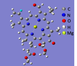
Figure 01: Geometrically optimized structure of Chlorophyll B molecule
The Chlorophyll-b molecule structure consists of a nitrogen- containing Mg at the center of molecules surrounded by four nitrogen atoms structure called a porphyrin ring; attached to the ring is a long carbon- hydrogen side chain Known as phytol chain [5]. For our simplicity phytic tail is removed. Chlorophyll is the primary photosynthetic pigment in green plants for the transfer of light energy to chemical acceptor [6]. Light that is absorbed provides the energy for photosynthesis. A green leaf absorbs blue light (mostly at 430nm) and red light (mostly at 600nm), and reflects back the green light, appearing green to human eyes [6]. Chlorophyll a, alone is found in blue-green and some red algae. Accessory pigment in Photosynthesis transfer light energy to chlorophyll a, one of these is chlorophyll b found in higher plants and green algae with chlorophyll a [7]. Chlorophyll c is also an accessory pigment found with Chlorophyll a in brown algae and diamonds [8]. Chlorophyll d, together with chlorophyll a, is in some red algae. Chlorophyll, except c, has ‘head’ and long ‘tail’ [9]. The head consists of a porphyrin ring of tetrapyrrole nucleus, from which extends a tail made up of a 20- carbon grouping called the phytol [10]. The defining elements of a chlorophyll molecule is the magnesium ion at the centre of a chlorine ring which make it ionic and hydrophilic and a ring is hydrophobic in nature with a carbonyl group which makes it polar [11]. Chlorophyll molecule undergoes change in their electronic states as that of bacteriochlorophyll-a, pheophytin, QA and QB molecule upon cation formation. One can extract the accurate vibrational information about molecules by FTIR difference spectroscopy [12]. In our research we have calculated FTIR difference spectra of labeled and unlabeled chlorophyll molecule. FTIR difference spectra obtained using the non-labeled and labeled particles allowed us to purpose assignments for many of the bands in difference spectra [13]. To calculate vibrational properties of neutral and cation forms of a model chlorophyll B molecule that is unlabeled, fully deuterated, 15 N, 18O, 13C and 2H and to gain some insights into what types of isotope induced frequency shifts might be expected for chlorophyll molecule we have used density functional theory DFT. We can directly compare the calculated isotope-induced shifts to experimentally observed band shift in the FTIR difference spectra [12]. If a direct connection between theory and experiment can be established then this will set a firm foundation for studies on more complex system [14].
- MATERIALS AND METHODS
Computational modes are mathematical models used to numerically study the behavior of complex system by means of a computer simulation. This method is used to predict the system’s behaviors under different conditions often for case in which instructive analytical solutions are not available. Geometrical Optimization and vibrational mode analysis of chlorophyll molecule were performed using hybrid density functional theory (DFT) [15]. In order to study the details of bio-molecules we have applied computational method using software GAUSSIAN 09W [16]. All stereo-chemical properties such as energy, bond length, frequency, dihedral angle etc. can be calculated from GAUSSIAN 09W. By using hybrid DFT method within Gaussian 09W geometrical optimization and IR absorption vibration modes of frequency calculations were done. The functional method B3LYP along with basis set 631-G (d) were employed for all the calculations. The molecule is optimized and different labeling of isotopes of atoms of chlorophyll molecules is calculated throughout this study.
- RESULTS AND DISCUSSIONS
We have studied the various isotopes labeling of elements in chlorophyll b molecule using Gaussian 09W in gas phase. For first of all we set the dihedral angle in the position of 3C of C2H3 group and rotate the dihedral angle from 0 degree to 360 degree in the interval of 10 degree. Afterwards we calculated the corresponding energy values by using Gaussian 09w on the 6-31G basis set from which graph between the energy along y-axis and angle along x-axis was plotted. The different possible energy values at different angles were noted among which the minimum value was obtained at 30 degree in Neutral state. In our research, we have discussed only the most intense vibrational modes lying in1900-1400cm-1. Here we have calculated the FTIR difference spectra of labeled and unlabeled isotopes of different atoms of chlorophyll- b molecule. In a Gauss view structure of chlorophyll-b molecules we replaced 12C to 13C isotopes, 16O to 18O isotopes, 14N to 15N isotopes with the help of word pad and gauss View 5.0 at temperature 98.18K and pressure 1 atmosphere and then calculated the vibrational frequency and intensity at different minimum energy, using density functional theory (DFT) with B3LYP functional and the 6-31G(d) basis set in gas phase.
3.1 Vibrational Properties of unlabeled and labeled Oxygen isotope (18O) of Chlorophyll-B molecule in Neutral state
Labeling of Oxygen isotope (18O) in Chlorophyll-B molecule has 237 normal modes of vibration. Most of these modes have very low infrared intensities and are undetectable in FTIR absorbance spectrum. For this reason, we have discussed only the most intense vibrational modes lying in 1900-1400 cm-1. Calculated harmonic Vibrational modes frequencies and intensities for the most prominent modes of unlabeled and labeled Oxygen isotope (18O) of Chlorophyll B are presented in Table 1. Figure below shows the calculated IR absorbance Spectra for unlabeled and labeled oxygen (18O) isotope as well as their corresponding difference for chlorophyll B molecule in 1900-1400cm-1 region. No negative frequency of chlorophyll B molecules is found while calculating frequency using density functional theory with B3LYP functional and the 6-31G (d) basis set in gas phase for both unlabeled and labeled state indicating that the molecule studied is the true energy minimized structure.
Table 01: Prominent harmonic vibrational mode frequencies and intensities calculated for unlabeled and labeled Oxygen isotope (18O) of Chlorophyll-B molecule. Mode assignments are also listed. Frequencies are in cm-1.
|
Modes |
Unlabeled Chl-b
(angle 300) ( freq cm-1) |
Labeled Chl- b(18O)
(angle 300) ( freq cm-1) |
| 173-ester C=O vibration | 1815.71 (202.12) | 1781.40 (194.62) |
| 131-keto C=O vibration | 1803.40 (603.49) AS
1811.19 (166.86) S |
1776.70 (166.86) AS
1769.29 (576.65) S |
| 71-keto vibration | 1758.81 (327.45) | 1727.81 (307.006) |
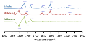
Figure 02: Calculated IR absorption spectra of Chl-b in gas phase. The labeled (upward) and unlabeled (middle) and their difference (downward) of Oxygen isotopic IR spectra are shown
From above table, it is observed that the 131-keto C=O mode have both symmetric and anti-symmetric mode of vibrational frequency. Symmetric mode occurs at 1811.19 cm-1and downshifted by 41.9 to 1769.29cm-1 upon labeled condition whereas anti-symmetric mode occurs at 1803.40 cm-1 and downshifted by26.7 to 1776.70 cm-1 at labeled condition. The 131-keto mode decreases in intensity by 436.63 km/mol (603.49 to 166.86) in anti-symmetric mode where as in symmetric mode the intensity increases by 409.79km/mol (166.86 to 576.65). The calculated 173-ester C=O stretching mode of chlorophyll at neutral state occurs at 1815.71 cm-1 and downshifted by 34.31 to 1781.40 cm-1 upon labeled condition. Intensity of this mode decreases by 7km/mol (202.12 to 194.62). Similarly, The calculated 71-keto C=O stretching mode occurs at 1758.81 cm-1 and downshifted by 31 to 1727.81 cm-1. The intensity of 71-keto mode decreases by 20.44 km/mol (327.45 to 307.006) upon cation formation.
Table 02: Summary of frequency and intensity of Unlabeled and labeled Oxygen isotope (18O) of neutral Chlorophyll – B molecules induced frequency shift of 131– keto C=O, 173-ester C=O and 131-keto modes.
| Molecule | 131 – keto mode | 173 – ester mode | 71– keto mode |
| Neutral Chl-b molecule | Symmetric = -41.9 (409.79)
Anti -symmetric = -26.7 ( -436.63) |
-34.31 (-7.5) | -31(-20.44) |
3.2 Vibrational Properties of unlabeled and labeled Carbon Isotope (13C) of Chlorophyll-B molecule in Neutral state
Labeling of Carbon isotope (13C) in Chlorophyll-B molecule has 237 normal modes of vibration. Most of these modes have very low infrared intensities and are undetectable in an FTIR absorbance spectrum. For this reason, we have discussed only the most intense vibrational modes lying in 1900-1400 cm-1. Calculated harmonic vibrational modes frequencies and intensities for the most prominent modes of unlabeled and labeled Carbon isotope (13C) of Chlorophyll B are presented in Table 3. Figure below shows the calculated IR absorbance Spectra for unlabeled and labeled carbon (13C) isotope as well as their corresponding difference for chlorophyll B molecules in 1900-1400 cm-1 region. No negative frequency of chlorophyll B molecule is found while calculating frequency using density functional theory with B3LYP functional and the 6-31G (d) basis set in gas phase for both unlabeled and labeled state indicating that the molecule studied is the true energy minimized structure.
Table 03: Prominent harmonic vibrational mode frequencies and intensities calculated for unlabeled and labeled Carbon isotope (13C) of Chlorophyll-B molecule. Mode assignments are also listed. Frequencies are in cm-1.
| Modes | Unlabeled Chl-b
(angle 300) ( freq cm-1) |
Labeled Chl- b (13C)
(angle 300) ( freq cm-1) |
| 173– ester C=O vibration | 1815.71 ( 202.12) | 1770.44 (190.00) |
| 131– keto C=O vibration | 1803.40 (603.49) AS
1811.19 (166.86) S |
1757.86 (592.69)AS
1765.63 (137.42) S |
| 71– keto vibration | 1758.81 (292.45) | 1715(309.95) |
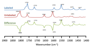
Figure 03: Calculated IR absorption spectra of Chl-b in gas phase. The labeled (upward) and unlabeled (middle) and their difference (downward) of Carbon isotopic IR spectra are shown
In this case, single vibration of 131-keto and 133 ester C=O mode vibration get mixed and the 131-keto C=O mode has major contribution to the anti-symmetric mode of vibration at 1803.40 cm-1 and 131-keto mode has major contribution to the symmetric mode of vibration at 1811.19 cm-1. The symmetric stretching of C=O mode of IR intensity is higher for Unlabeled than that for labeled. Anti-symmetric 131-keto C=O vibration of Unlabeled molecule occurs at 1803.40 cm-1 whereas in labeled carbon isotope 13C, it is downshifted by 45.54 and occurs at 1757.86 cm-1. Intensity in this mode is decreased by 10.8 km/mol (603.49 to 592.69) upon labeling. Symmetric 131-keto vibration of unlabeled carbon occurs at 1811.19 cm-1 and downshifted by 46.56 to 1765.63 cm-1 for labeled carbon isotope (13C). In this case, the intensity is decreased by 29.44 km/mol (166.86 to 137.42) upon labeled condition. In unlabeled carbon isotope, the stretching of 71-keto mode of IR intensity of unlabeled mode occurs at1758.81 cm-1 which is downshifted by 43.81 to 1715 cm-1 upon labeled conditions. Intensity in this condition is decreased by 17.5 km/mol (327.45 to 309.95) under labeling. Also, Unlabeled 173 ester C=O mode of IR intensity occurs at 1815.71 cm-1 and labeled carbon isotopes 13C is decreased by 45.27 and occurs at 1770.44 cm-1 whereas intensity is decreased by 12.12 km/mol (202.12 to 190.00) at Neutral mode.
Table 04: Summary of frequency and intensity of Unlabeled and labeled Carbon Isotope (13C) of neutral Chlorophyll – B molecules induced frequency shift of 131– keto C=O, 173– ester C=O and 71-keto modes.
| Molecule | 131– keto | 173– ester | 71-keto |
| Neutral Chl-b molecule | Anti-symmetric= – 45.54(-10.8)
Symmetric = – 46.56 (-29.44) |
– 45.27(-12.12) | – 43.81(-17.5) |
3.3 Vibrational Properties of unlabeled and labeled Nitrogen Isotope (15N) of Chlorophyll-B molecule in Neutral state
Labeling of Nitrogen isotope (15N) in Chlorophyll-B molecule has 237 normal modes of vibration with most of these having very low infrared intensities. Thus, only the most intense vibrational modes lying in 1900-1400 cm-1 is discussed. Calculated harmonic vibrational modes frequencies and intensities for the most prominent modes of unlabeled and labeled Nitrogen isotope (15N) of Chlorophyll B are presented in Table 5. Figure below shows the calculated IR absorbance Spectra for unlabeled and labeled Nitrogen (15N) isotope as well as their corresponding difference for chlorophyll B molecules in 1900-1400 cm-1 region. No negative frequency of chlorophyll B molecule is found while calculating frequency using density functional theory with B3LYP functional and the 6-31G (d) basis set in gas phase for both unlabeled and labeled state indicating that the molecule studied is the true energy minimized structure.
Table 05: Prominent harmonic vibrational mode frequencies and intensities calculated for unlabeled and labeled Nitrogen isotope (15N) of Chlorophyll-B molecule. Mode assignments are also listed. Frequencies are in cm-1.
| Modes | Unlabeled Chl-b
(angle 300) ( freq cm-1) |
Labeled Chl-b (15N)
(angle 300) ( freq cm-1) |
| 173-ester C=O vibration | 1815.71 (202.12) | 1815.71 (202.26) |
| 131-keto C=O vibration
|
1803.40 (603.49) AS
1811.19 (166.86) S |
1803.37 (605.94)AS
1811.16 (165.63)S |
| 71– keto vibration | 1758.81(327.45) | 1758.80 (327.68) |
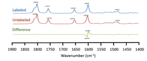
Figure 04: Calculated IR absorption spectra of Chl-b in gas phase. The labeled (upward) and unlabeled (middle) and their difference (downward) of Nitrogen isotopic IR spectra are shown
In this case, single vibration of 131-keto and 133 ester C=O mode vibration get mixed and the 131-keto C=O mode has major contribution to the anti-symmetric mode of vibration at 1803.40 cm-1 and 131-keto mode has major contribution to the symmetric mode of vibration at 1811.19 cm-1. The symmetric stretching of C=O mode of IR intensity is higher at Unlabeled and labeled. Anti-symmetric 131-keto C=O vibration of Unlabeled groups occurs at 1803.40 cm-1 whereas in labelled carbon isotopes 13C, it is nearly same as it is downshifted by 0.3 occurs at 1803.37 cm-1. The intensity in this case is slightly increased by 0.14 km/mol (202.12 to 202.26). In case of symmetric 131-keto, the modes of vibration occurs at 1811.19 cm-1 and nearly remains unchanged since it is downshifted by 0.3 and occurs at 1811.16 cm-1. Intensity in this case is increased by 1.22 km/mol (164.41 to 165.63). 173– ester vibration of unlabeled Nitrogen occurs at 1815.71 cm-1 and remains unchanged at labeling conditions whereas intensity in this case remains nearly unchanged as it is slightly increased by 0.23 km/mol (327.45 to 327.68).
Table 06: Summary of frequency and intensity of Unlabeled and labeled Nitrogen Isotope (15N) of neutral Chlorophyll-B molecules induced frequency shift of 131– keto C=O, 173– ester C=O and 131-keto modes.
| Molecule | 131– keto | 173– ester | 131-keto |
| Neutral Chl- b molecule | Anti-symmetric = – 0.03(2.45)
Symmetric = – 0.03(1.22) |
Unchanged. |
– 0.01(0.23) |
ACKNOWLEDGMENT
We are grateful to the Physics Department of St. Xavier’s College for providing us Computational lab for out computational work, and all the team members for their hard work to accomplish this task
REFERENCES
- N. Glazer, Structure and molecular organization of the photosynthetic accessory pigments of cyanobacteria and red algae, Mol. Cell. Biochem. 18 (1977) 125–140.
- R. Acharya, P. Lamichhane, R. Wahab, D.K. Chaudhary, B. Shrestha, L.P. Joshi, N.K. Kaushik, E.H. Choi, Study on the Synthesis of ZnO Nanoparticles Using Azadirachta indica Extracts for the Fabrication of a Gas Sensor, Molecules. 26 (2021) 7685.
- P. Lamichhane, Calculated vibrational properties of quinones in photosynthetic reaction centers, (2011).
- B. Borkar, M. Negi, A. Jaiswal, T.R. Acharya, N. Kaushik, E.H. Choi, N.K. Kaushik, Plasma-Generated Nitric Oxide Water: A promising strategy to combat bacterial dormancy (VBNC state) in environmental contaminant Micrococcus luteus, J. Hazard. Mater. (2023) 132634.
- Sumanta, C.I. Haque, J. Nishika, R. Suprakash, Spectrophotometric analysis of chlorophylls and carotenoids from commonly grown fern species by using various extracting solvents, Res J Chem Sci. 2231 (2014) 606X.
- HILL, Chlorophyll, in: Compr. Biochem., Elsevier, 1963: pp. 73–97.
- W. Qiu, D.C. Jiang, X.S. Wang, B.S. Wang, F. Zhou, Advances in the members and biosynthesis of chlorophyll family., Photosynthetica. 57 (2019).
- Rakin Hossain Rayean & Narayan Chandra Nath (2023). Design of Solar Cell with Comparative Analysis on Different Parameter Using PSpice. Dinkum Journal of Natural & Scientific Innovations, 2(12):750-764.
- J. Hibberd, The structure and phylogenetic significance of the flagellar transition region in the chlorophyll c-containing algae, BioSystems. 11 (1979) 243–261.
- L. \.Inanç, Chlorophyll: Structural Properties, Health Benefits and Its Occurrence in Virgin Olive Oils., Acad. Food Journal/Akademik GIDA. (2011).
- Scheer, Structure and occurence of chlorophylls, (1991).
- M.P. Bandaranayake, V. Sivakumar, R. Wang, G. Hastings, Modeling the A1 binding site in photosystem: I. Density functional theory for the calculation of “anion- neutral” FTIR difference spectra of phylloquinone, Vib. Spectrosc. 42 (2006) 78–87.
- Sivakumar, R. Wang, G. Hastings, A1 reduction in intact cyanobacterial photosystem I particles studied by time-resolved step-scan Fourier transform infrared difference spectroscopy and isotope labeling, Biochemistry. 44 (2005) 1880–1893.
- Wang, S. Parameswaran, G. Hastings, Density functional theory based calculations of the vibrational properties of chlorophyll-a, Vib. Spectrosc. 44 (2007) 357–368.
- J. Frisch, G.W. Trucks, H.B. Schlegel, G.E. Scuseria, M.A. Robb, J.R. Cheeseman, J.A. Montgomery Jr, T. Vreven, K.N. Kudin, J.C. Burant, others, Gaussian 03, Revision C. 02. Wallingford, CT: Gaussian, Inc.[Google Sch. (2004).
- R. Rijal, H.P. Lamichhane, K. Pudasainee, Molecular structure, homo-lumo analysis and vibrational spectroscopy of the cancer healing pro-drug temozolomide based on dft calculations, AIMS Biophys. 9 (2022) 208–220.
Publication History
Submitted: December 22, 2023
Accepted: January 14, 2024
Published: January 31, 2024
Identification
D-0211
Citation
Prabesh Adhikari, Suresh Khadka, Laxmi Prasad Odari, Yamnath Poudel, Sandhya Gautam & Hari Prasad Lamichhane (2024). Computational Study on Vibrational Properties of Chlorophyll-B Model Molecules in Gases Phases. Dinkum Journal of Natural & Scientific Innovations, 3(01):101-108.
Copyright
© 2024 DJNSI. All rights reserved