
Vol. 5 No.02 (2026) | Page: 190-199 |Case Study 
Closing the Neurological Safety Gap: A Case Study of a Rapid MRI Pathway for Suspected Cauda Equina Syndrome
Narikazu Boku 1*
-
- Institute of Medical Science, The University of Tokyo, Tokyo, Japan.
 nboku@ims.u-tokyo.ac.jp




ABSTRACT
Background: Cauda equina syndrome (CES) is a time-critical spinal emergency caused by compression of the lumbosacral nerve roots. Delayed diagnosis and MRI can lead to permanent bladder, bowel, sexual, sensory, and lower-limb dysfunction. Recent guidance emphasizes rapid assessment and 24/7 MRI access for suspected cases.
Objective: To examine system failures contributing to delayed CES diagnosis and propose pathway improvements that support timely imaging and intervention.
Methods: TA composite hospital case involving a patient with progressive back pain, bilateral radicular symptoms, saddle sensory changes, and urinary disturbance was analyzed. Evidence-based CES service recommendations were reviewed to identify opportunities for pathway redesign.
Results: The case demonstrated avoidable delays before MRI, increasing the risk of neurological deterioration. Key interventions included direct emergency department MRI requests, standardized red-flag documentation, CES checklists, radiology escalation protocols, audit monitoring, and structured post-imaging surgical pathways.
Conclusion: Effective management of suspected CES depends on both clinical expertise and reliable systems. CES should be treated as a pathway emergency requiring rapid imaging, coordinated multidisciplinary care, and streamlined referral processes to minimize permanent neurological disability.
Keywords: cauda equina syndrome, spine surgery, neurology, MRI pathway, spinal emergency
Key points:
- Can streamlined emergency pathways reduce diagnostic delays and prevent neurological deterioration in patients with suspected cauda equina syndrome?
- Avoidable MRI delays increase the risk of neurological deterioration and potentially permanent disability in patients with suspected cauda equina syndrome.
- Rapid MRI access, standardized red-flag documentation, and coordinated multidisciplinary referral are essential to prevent permanent neurological disability.
- INTRODUCTION
Cauda equina syndrome (CES) is a rare but potentially catastrophic spinal emergency caused by compression of the lumbosacral nerve roots, most commonly from a large lumbar disc herniation. Its clinical significance lies less in its incidence than in the severity and permanence of harm that can follow delayed recognition, including bladder and bowel dysfunction, sexual dysfunction, saddle sensory loss, lower-limb weakness and chronic neuropathic disability [1,2]. Consequently, suspected CES represents a high-stakes diagnostic problem in which neurological safety depends on rapidly distinguishing the relatively small proportion of patients with compressive pathology from the much larger group presenting with clinically similar symptoms. This distinction is difficult at the bedside. Traditional “red-flag” symptoms including urinary disturbance, saddle anesthesia, bilateral sciatica and reduced anal tone have limited diagnostic accuracy when considered individually, and systematic evidence demonstrates substantial heterogeneity in their sensitivity and specificity for MRI-confirmed CES [3]. More recent reviews similarly conclude that no history or physical-examination feature can reliably exclude clinically important cauda equina compression and that suspected cases therefore require urgent radiological investigation [4]. This creates an unavoidable safety–efficiency tension: restrictive imaging risks a devastating missed diagnosis, whereas indiscriminate emergency referral generates large numbers of negative MRI examinations, additional transfers, prolonged emergency-department stays and pressure on already constrained radiology services.Magnetic resonance imaging (MRI) therefore functions as the decisive diagnostic gateway between clinical suspicion and definitive spinal management. UK Clinical Imaging Board guidance recommends access to 24-hour MRI for acute suspected CES and states that imaging should be undertaken as soon as practicable and within four hours of the request, with the final report available rapidly thereafter [5]. The updated National Suspected Cauda Equina Syndrome Pathway reinforces this position, identifying MRI at the presenting hospital as best practice and maintaining the expectation that emergency imaging should occur as soon as possible and certainly within four hours [6]. Importantly, adjunctive tests should not become barriers to imaging: post-void residual bladder volume can support assessment, but a value below 200 mL does not reliably exclude incomplete CES, and MRI remains necessary where clinical suspicion persists [6,7]. The principal neurological safety gap is therefore increasingly organisational rather than technological. MRI is well established as the investigation of choice, yet access may still depend on time of presentation, local scanner availability, radiographer staffing, reporting capacity, referral processes and inter-hospital transfer arrangements. A two-centre UK study demonstrated the inefficiency of transferring suspected cases for out-of-hours imaging: among 339 transferred patients, only 16 (4.7%) demonstrated cauda equina compression requiring urgent decompression, while half of those with negative scans experienced discharge or repatriation delays exceeding 24 hours [8]. Such findings indicate that conventional referral pathways can simultaneously expose true CES cases to avoidable delay and subject scan-negative patients to unnecessary movement and prolonged hospital utilisation. Rapid or direct-to-MRI pathways offer a service-level response to this problem by redesigning the diagnostic sequence around timely imaging rather than sequential specialty gatekeeping. Recent UK service evaluations demonstrate growing interest in pathways that enable patients with appropriately screened CES symptoms to progress rapidly from community or emergency assessment to definitive MRI investigation [9]. Quality-improvement evidence further suggests that achieving a four-hour request-to-scan target alone may be insufficient when delays occur earlier in the patient journey, highlighting the importance of examining the whole pathway from presentation to imaging, reporting and specialist decision-making [10].Against this background, the present case study evaluates a rapid MRI pathway for suspected CES as a neurological patient-safety intervention rather than simply a radiology-access initiative. The central concern is whether pathway redesign can reduce diagnostic delay while maintaining appropriate clinical selection, efficient resource utilisation and timely escalation of MRI-positive patients. By examining the interaction between clinical recognition, referral, imaging access, reporting and onward spinal management, the case study addresses the critical question of how healthcare systems can close the gap between national standards for time-critical CES diagnosis and the realities of frontline service delivery.
- CASE PRESENTATION
A 47-year-old warehouse worker presented to the emergency department (ED) with severe progressive lower-back pain radiating into both legs. The symptoms had deteriorated over several days and were associated with new neurological complaints, including numbness affecting the inner thighs/perineal region and difficulty initiating urination. The patient had no previous history of spinal surgery or known spinal pathology. At initial triage, the presenting complaint was documented as “back pain with sciatica”, and symptomatic analgesic treatment was commenced. Due to significant emergency department workload, the patient remained awaiting further clinical assessment before senior review. Although back pain with radiculopathy is a common ED presentation, the combination of bilateral neurological symptoms and urinary disturbance represented potential warning signs of evolving cauda equina syndrome (CES).
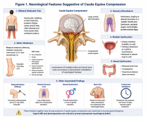
Figure 01: Neurological Features Suggestive of Cauda Equina Compression.
Figure 01. Representative illustration demonstrating the anatomical distribution of cauda equina nerve roots and the saddle sensory area affected in CES. The development of altered perineal sensation and urinary difficulty indicated possible involvement of the lower sacral nerve roots responsible for bladder, bowel and sensory function. These symptoms increased the clinical concern from uncomplicated lumbar radiculopathy to a time-critical neurological emergency requiring urgent investigation.
2.1 Senior Clinical Assessment
During subsequent assessment by the emergency clinician, the patient was identified as having several features concerning for CES: bilateral radicular leg symptoms; altered perineal sensation by history; and new urinary dysfunction characterised by difficulty initiating urination. A neurological examination was performed, and a post-void bladder assessment was requested. Although bladder scanning can provide additional information regarding urinary dysfunction, it cannot independently exclude CES. Therefore, the clinical presentation required definitive spinal imaging. The patient was considered to have possible incomplete cauda equina syndrome, where neurological compromise is developing but complete urinary retention has not yet occurred. Early recognition at this stage is important because irreversible neurological injury may occur if decompression is delayed.
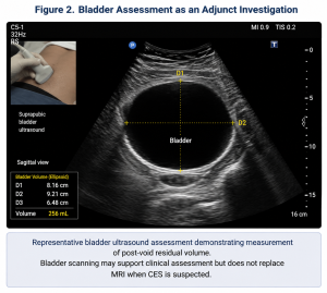
Figure 02: Bladder Assessment as an Adjunct Investigation
Figure 02 representative bladder ultrasound assessment demonstrating measurement of post-void residual volume. Bladder scanning may support clinical assessment but does not replace MRI when CES is suspected.The use of bladder assessment highlights an important aspect of CES evaluation: supportive investigations can improve clinical decision-making, but urgent MRI remains the definitive diagnostic investigation when neurological symptoms suggest possible nerve-root compression.
2.2 Diagnostic Pathway and Delay Points
Following recognition of possible cauda equina syndrome (CES), the emergency clinician-initiated arrangements for urgent lumbar MRI. However, the local diagnostic pathway required several sequential steps before imaging could be undertaken, including discussion with the on-call specialty team, documentation of detailed neurological examination findings, confirmation that the patient met the criteria for suspected CES imaging, and radiology approval for MRI access. Consequently, the patient remained in the emergency department while awaiting definitive imaging. This sequence highlights the central neurological safety gap identified in this case: although CES was clinically recognized and urgent imaging was considered necessary, organizational and procedural requirements introduced additional stages between initial clinical suspicion and diagnostic confirmation, creating potential opportunities for delay in a time-critical neurological condition.
2.3 MRI Findings
Emergency MRI of the lumbar spine was eventually performed and demonstrated a large central lumbar disc herniation causing significant compression of the cauda equina within the spinal canal.
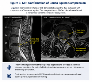
Figure 03: MRI Confirmation of Cauda Equina Compression
Figure 03 represents lumbar MRI demonstrating central disc extrusion with compression of the cauda equina. This image is from published clinical material and is not derived from the composite case patient. The MRI findings confirmed the suspected diagnosis and provided anatomical evidence explaining the patient’s bilateral radicular symptoms, sensory disturbance and urinary dysfunction. The transition from suspected CES to confirmed structural compression allowed urgent spinal surgical decision-making.
2.4 Surgical Management and Outcome
Following MRI confirmation, the spinal surgical team accepted the patient for urgent decompression of the affected lumbar level. The aim of surgery was to relieve pressure on the compressed nerve roots and prevent further neurological deterioration. Following surgery, the patient demonstrated significant improvement in bilateral leg pain. However, urinary symptoms showed only partial recovery during early follow-up. This outcome reflects an important clinical principle in CES management: although decompression removes the mechanical cause of nerve compression, recovery depends on the severity and duration of neurological injury before treatment. Delayed diagnosis may therefore result in persistent bladder, bowel or sensory impairment despite technically successful surgery.
2.5 Pathway Analysis: Identifying the Neurological Safety Gap
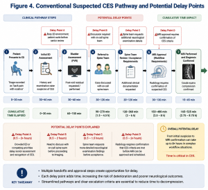
Figure 04: Conventional Suspected CES Pathway and Potential Delay Points
The case demonstrates that the major challenge in suspected CES is not simply availability of MRI technology, but the efficiency of the pathway connecting clinical recognition to definitive diagnosis. A rapid MRI pathway aims to reduce avoidable delays by allowing appropriately trained clinicians to directly activate urgent imaging when CES criteria are met, while maintaining specialist involvement after imaging confirmation.
2.5 Case Interpretation
This composite case illustrates how delays in suspected CES management can occur despite appropriate clinical awareness. The patient’s neurological symptoms were eventually recognised, MRI confirmed cauda equina compression, and urgent decompression was undertaken. However, the period between suspicion and imaging represented a critical window during which neurological deterioration could occur. The case supports the development of streamlined rapid MRI pathways that minimise unnecessary administrative barriers, improve coordination between emergency medicine, radiology and spinal services, and prioritise time-sensitive neurological diagnosis.
2.6 Clinical and System Problem
The central clinical problem was possible compression of the lumbosacral nerve roots with evolving neurological dysfunction. The system problem was delayed movement from suspicion to imaging. BMJ Open Quality describes suspected cauda equina syndrome as a surgical emergency requiring prompt MRI diagnosis and surgical treatment to prevent permanent bowel and bladder incontinence, sexual dysfunction, lower-limb paralysis, and chronic pain. The same article reports that the GIRFT National CES Pathway 2023 mandates emergency MRI “as soon as possible, certainly within 4 hours of request.” The Royal College of Radiologists guidance also sets service standards for MRI provision, including timing, referral criteria, access, staffing, reporting, protocols, and audit. In this case, multiple safeguards existed but were not connected. Clinicians knew cauda equina syndrome was serious. Radiology had MRI capability. Spine surgeons were available for consultation. Yet the patient pathway moved slowly because responsibility was fragmented among triage, emergency review, specialty referral, MRI approval, imaging, reporting, and surgical acceptance. The failure was not a single missed diagnosis. It was a system that did not convert suspicion into immediate diagnostic action.
2.7 Intervention Design
The hospital convened a multidisciplinary spine-neurology safety group. Membership included emergency physicians, spine surgeons, neurologists, radiologists, radiographers, emergency nurses, bed managers, and quality improvement staff. The group’s aim was to reduce delays from emergency department arrival to MRI completion for patients with suspected cauda equina syndrome.
The pathway redesign had six components.
- Direct emergency department MRI requesting
Emergency clinicians were authorized to request suspected cauda equina MRI directly when documented red-flag symptoms were present. This reduced dependency on sequential approval from multiple teams.
- Standardized red-flag documentation
A suspected cauda equina template was embedded into the emergency department record. It prompted documentation of bilateral radicular pain, saddle or perineal sensory change, bladder disturbance, bowel disturbance, lower-limb weakness, sensory deficit, and relevant examination findings.
- Radiology escalation protocol
MRI requests labeled “suspected cauda equina syndrome” triggered direct notification to the radiology coordinator and on-call reporting radiologist. If MRI could not be performed on site within the expected pathway window, escalation to the duty manager and spine team was required.
- MRI operational planning
The radiology department mapped scanner availability, staffing gaps, and after-hours constraints. The Royal College of Radiologists guidance identifies 24/7 MRI access, staffing, radiographer training, image transfer, reporting, protocol, and audit as service standards for suspected cauda equina imaging.
- Surgical decision pathway
MRI results were categorized as: confirmed cauda equina compression, alternative urgent spinal pathology, non-compressive findings, or indeterminate imaging requiring senior review. Confirmed compression triggered immediate spine surgical discussion.
- Audit and feedback
Monthly audits reviewed time from emergency arrival to MRI request, request to MRI completion, MRI completion to report, report to spine decision, and final disposition. The BMJ Open Quality report emphasized that even when request-to-MRI targets are met, delays can still occur from emergency department attendance to MRI scan. Therefore, the hospital measured the whole pathway rather than only the imaging interval.
- DISCUSSION
This case illustrates a common challenge in spine-neurology emergencies: knowledge is necessary but insufficient. Most clinicians understand that cauda equina syndrome is serious. The problem is that real-time hospital systems often make urgent action difficult. In suspected cauda equina syndrome, MRI is not simply a diagnostic test; it is the gateway to neurological decision-making. If MRI is delayed, surgical decision-making is delayed. If referral criteria are unclear, imaging may be delayed. If red flags are not documented consistently, radiology and surgical teams may hesitate. If MRI is available only during limited hours, timely decision-making becomes dependent on the clock rather than the patient’s neurological risk.The case also shows why whole-pathway measurement is essential. A hospital can meet an imaging target measured from request time but still fail patients if the request itself is delayed. A journal article reported that the Medway Hospital project identified time to MRI request as a major driver of delays, leading the team to change its primary aim to MRI within 4 hours from emergency department presentation rather than from request. This change reflects a crucial principle: patients experience the pathway from arrival, not from the moment a form is submitted. The case also has implications for neurology and spine education. Frontline clinicians should be trained to recognize that cauda equina syndrome may present with evolving symptoms. Documentation should not be limited to pain severity. Bladder function, bowel function, saddle sensation, bilateral symptoms, motor findings, sensory findings, and gait should be actively assessed and recorded.Finally, the case highlights the importance of balancing sensitivity and specificity. Because suspected cauda equina syndrome has serious consequences, pathways should support timely imaging for appropriate red-flag presentations. At the same time, indiscriminate MRI use can overwhelm radiology services. Standardized criteria and audit can help identify patients who need urgent imaging while reducing avoidable scans.
- CONCLUSION
Cauda equina syndrome is a neurological emergency in which delay can permanently alter a patient’s life. This case study shows that the central challenge is not only diagnostic knowledge, but pathway reliability. Hospitals must ensure that suspected cauda equina syndrome triggers rapid assessment, structured documentation, urgent MRI, timely reporting, and immediate spine surgical decision-making. The broader lesson for spine and neurology services is clear: neurological outcomes depend on systems as much as specialists. A safe pathway is one in which the right patient receives the right scan, at the right time, with the right team ready to act.
AUTHOR CONTRIBUTION STATEMENT
Narikazu Boku: Substantial contributions to the conception or design of the work; or the acquisition, analysis, or interpretation of data for the work, Drafting the work or reviewing it critically for important intellectual content; AND Final approval of the version to be published, and Agreement to be accountable for all aspects of the work in ensuring that questions related to the accuracy or integrity of any part of the work are appropriately investigated and resolved.
ACKNOWLEDGEMENTS: No specific individuals, institutions, or organizations contributed additional support or assistance requiring formal acknowledgement for this study.
DECLARATIONS:
Funding: This research received no specific grant, financial support, or funding from any public, commercial, or non-profit organization.
Conflicts Of Interest: The authors declare that there are no conflicts of interest, financial or otherwise, that could have influenced the research, analysis, or interpretation of the findings presented in this article.
Data Availability: The datasets and information used in this study are publicly available from the sources cited within the article. No proprietary or restricted data were used.
Trial Registration: Trial registration is not applicable to this study, as it does not involve clinical trials, human participants, or experimental interventions.
Generative AI Disclosure: Generative artificial intelligence tools were not used for generating research data, conducting analyses, or producing scientific conclusions in this study. The authors take full responsibility for the content and accuracy of the manuscript.
REFERENCES
- Lavy C, Marks P, Dangas K, Todd N. Cauda equina syndrome—a practical guide to definition and classification. International Orthopaedics. 2022;46(2):165–169. doi:10.1007/s00264-021-05273-1.
- Hoeritzauer I, Wood M, Copley PC, Demetriades AK, Woodfield J. What is the incidence of cauda equina syndrome? A systematic review. Journal of Neurosurgery: Spine. 2020;32(6):832–841. doi:10.3171/2019.12.SPINE19839.
- Dionne N, Adefolarin A, Kunzelman D, et al. What is the diagnostic accuracy of red flags related to cauda equina syndrome when compared with magnetic resonance imaging? A systematic review. Musculoskeletal Science and Practice. 2019;42:125–133. doi:10.1016/j.msksp.2019.05.004.
- McNamee J, Flynn P, O’Leary S, Love M, O’Neill A, O’Byrne J. Imaging in cauda equina syndrome—a pictorial review. Insights into Imaging. 2013;4(6):669–676. doi:10.1007/s13244-013-0286-8.
- Fraser S, Roberts L, Murphy E. Cauda equina syndrome: a literature review of its definition and clinical presentation. Archives of Physiotherapy. 2009;1:1–7.
- Todd NV. Cauda equina syndrome: the timing of surgery probably does influence outcome. British Journal of Neurosurgery. 2011;25(5):589–592. doi:10.3109/02688697.2011.578568.
- Todd N, Dangas K, Lavy C. Post-void bladder ultrasound in suspected cauda equina syndrome—data from medicolegal cases and relevance to magnetic resonance imaging scanning. International Orthopaedics. 2022;46:1375–1380. doi:10.1007/s00264-022-05341-0.
- Silva A, Sachdev B, Kostusiak M, et al. Out of hours magnetic resonance imaging for suspected cauda equina syndrome: lessons from a comparative study across two centres. Annals of the Royal College of Surgeons of England. 2021;103(3):218–222. doi:10.1308/rcsann.2020.7086.
- Conte A, Lingham A, Nagulendran S, et al. Improving the suspected cauda equina syndrome pathway at a district general hospital: a quality improvement project. BMJ Open Quality. 2025;14:e003081. doi:10.1136/bmjoq-2024-003081.
- Clinical Imaging Board. MRI Provision for Cauda Equina Syndrome. London: Royal College of Radiologists, Society and College of Radiographers, Institute of Physics and Engineering in Medicine; 2023.
Supplemental Material
No supplementary files, datasets, figures, or additional materials are associated with this study. Any information necessary to understand and evaluate the work is included in the main article.
Citation
Narikazu Boku (2026). Closing the Neurological Safety Gap: A Case Study of a Rapid MRI Pathway for Suspected Cauda Equina Syndrome. Dinkum Journal of Medical Innovations, 5(03):190-199. https://doi.org/10.71017/djmi.5.03.d-0589
DOI
https://doi.org/10.71017/djmi.5.03.d-0589
Identification
D-0589
Publication History
Submitted: 21-01-2026
Accepted: 15-03-2026
Published: 31-03-2026
Copyright
© The Author(s) 2026
Publishing License

 This is an Open Access article distributed under the terms of the Creative Commons Attribution (CC BY 4.0) License .